Download
1 / 17
170 likes | 268 Vues
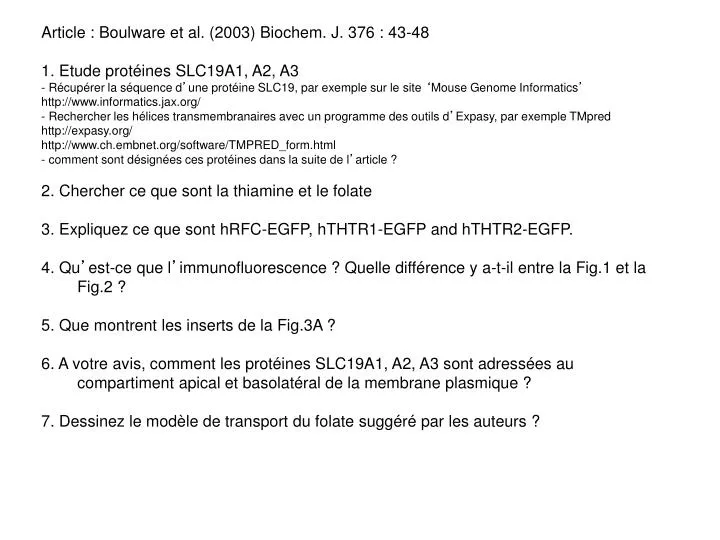
Article : Boulware et al. (2003) Biochem. J. 376 : 43-48 1. Etude protéines SLC19A1, A2, A3 - Récupérer la séquence d ’ une protéine SLC19, par exemple sur le site ‘ Mouse Genome Informatics ’ http://www.informatics.jax.org/

E N D
Article : Boulware et al. (2003) Biochem. J. 376 : 43-48 1. Etude protéines SLC19A1, A2, A3 - Récupérer la séquence d’une protéine SLC19, par exemple sur le site ‘Mouse Genome Informatics’ http://www.informatics.jax.org/ - Rechercher les hélices transmembranaires avec un programme des outils d’Expasy, par exemple TMpred http://expasy.org/ http://www.ch.embnet.org/software/TMPRED_form.html - comment sont désignées ces protéines dans la suite de l’article ? 2. Chercher ce que sont la thiamine et le folate 3. Expliquez ce que sont hRFC-EGFP, hTHTR1-EGFP and hTHTR2-EGFP. 4. Qu’est-ce que l’immunofluorescence ? Quelle différence y a-t-il entre la Fig.1 et la Fig.2 ? 5. Que montrent les inserts de la Fig.3A ? 6. A votre avis, comment les protéines SLC19A1, A2, A3 sont adressées au compartiment apical et basolatéral de la membrane plasmique ? 7. Dessinez le modèle de transport du folate suggéré par les auteurs ?
>P41438 P41438 mus musculus (mouse). folate transporter 1 (intestinal folate carrier protein) (ifc-1) (reduced folate carrier 1) (rfc-1) (rfc1) (solute carrier family 19 member 1). 6/2008 MVPTGQVAEKQAYEEPRQDHELKSWRCLVFYLCFFGFMAQLRPGESFITPFLLERKFTKE QVTNEIIPMLPYSHLAVLVPVFLLTDYLRYKPVLVLQCLSFVCVWLLLLLGTSVVHMQLM EVFYSVTMAARIAYSSYIFSLVHPSRYQRMASYSRAAVLLGVFISSVLGQALVTVGHIST YTLNCVSLGFILFSLVLSLFLKRPKRSLFFNRSTLARGALPCELDQMHPGPDRPETRKLD RMLGTCRDSFLVRMLSELVENARQPQLRLWCLWWVFNSSGYYLITYYVHVLWRSTDSSLS YNGAVDAASTLLSAITSFSAGFLSIRWTLWSKLVIAGVIAIQASLVFCMFQIRDIWVCYV TFVLFRGAYQFLVPIATFQIASSLSKELCALVFGINTFLATALKTCITLVVSDKRGLGLQ VRDQFRIYFIYFLMLSITCFAWAGLDGLRYCQRGRHQPLAQAQELRSPLETSVQAISLQD GDLRGPQPSAPQLLSEDGMEDDRGDLRVEAKA 1 2 3 4 5 6 7 8 9 11 12 10 SLC19A1 = RFC reduced folate carrier
>NP_473428 NP_473428 solute carrier family 19 (thiamine transporter), member 2 [Mus musculus]. 2/2008 MDVPARVSRRAAAAAARMLLRTARVPRECWFLPTALLCAYGFFANLRPSEPFLTPYLLGP DKNLTERQVYNEIYPVWTYSYLLLLFPVFLATDYLRYKPVILLQGLSLIVTWFMLLYAQG LLAIQFLEFFYGIATATEIAYYSYIYTVVDLGMYQKVTSYCRSATLVGFTVGSVLGQILV SVVGWSLFSLNVISLTCVSVAFAVAWFLPMPQKSLFFHHIPSSCHGVNGLKVQNGGIVTD TPAANHLPGWEDIESKIPLNLDEPPVEEPEEPKPDRLRVFRVLWNDFLMCYSSRPLLCWS VWWALSTCGYFQVVNYAQGLWEKVMPSQNADIYNGGVEAVSTLLGASAVFAVGYIKLSWS TWGEMTLFLCSLLIAAAVYVMDTVQSIWVCYASYVVFRIIYMVLITIATFQIAANLSMER YALVFGVNTFIALALQTLLTLIVVDARGLGLCITTQFLIYASYFAAISVVFLANGIVSII KKCRKQEDPSSSPQASTS 1 2 3 4 5 7 8 9 10 11 12 6 ? SLC19A2 = THTR1 thiamine transporter
>Q99PL8 Q99pl8 mus musculus (mouse). thiamine transporter 2 (thtr-2) (thtr2) (solute carrier family 19 member 3). 6/2008 MDSSCRTPPSNSWVYPTVILCLFGFFSMFRPSEAFLIPFLSEPSKNLTSPEMTNEILPVW TYSYLATLPPVFVLTDYLRYKPVIMLHVVAFATSYLFLLFGQGVMLMQTAEFFFGVVSAT EIAYFAYIYSMVSPEHYQKVSSYCRSITLVAYTAGSVLAQLLVSLTNLPYSSLFYISLAC VSVAFFFSLFLPMPKKSMFFHAKSDRDDCPKPLEQCTVPKEAQSNRTHSELFANSKNLED REMSNPDPENSALRHFAHWFQDLKECYSSKHLVYWSLWWAFATAGYNQILNYVQVLWEHK APSQDSSIYNGAVEAIATFGGALASFSVGYLKVNWDLLGELGLAVFSAVIAGSLFLMNYS RSIWVCYAGYLLVKSSYSFLITIAVFQIAVNLSLERYALVFGIDTFIALVIQTIMTMIVV DQRGLQLPVTTQFLVYGSYFAVIAGVFLMRSIYILCSAKCRKEVQNLATTRSPNEPHPQE PSNVSTKF 1 2 3 4 5 6 7 8 9 10 11 12 SLC19A3 = THTR2 thiamine transporter
Thiamine = vitamine hydrosoluble B1 Précurseur de la thiamine pyrophosphate, co-enzyme de carboxylases du métabolisme des glucides Besoin journalier : 1-1,6 mg Thiamine pyrophosphate Folate = vitamine hydrosoluble B9 Co-enzyme pour la synthèse des bases de l’ADN Besoin journalier : 200-800 µg Est réduit en DHF (dihydrofolate) et THF (tetrahydrofolate, forme active du folate). La réaction est catalysée par la dihydrofolate reductase (DHR) folate DHF DHR methotrexate folinic acid THF
Acide folinique : équivalent du tetrahydrofolate (forme active du folate). Ne se lie pas à la dihydrofolate reductase (DHR) Methotrexate : inhibiteur de la dihydrofolate reductase (DHR)
Microscopie de fluorescence Intensité de fluorescence émission excitation excitation émission Longueur d’onde
Quelques fluorophores courants Fluorophore lexlem Hydroxycoumarine 385 (UV)445 (bleu) Alexa 518 494 (bleu) 518 (vert) Fluorescéine* 494 (bleu)518 (vert) Rhodamine 570 (vert)590 (rouge) Alexa 590 570 (vert)590 (rouge) Cy 5 494 (bleu)518 (vert) Cy 3 570 (vert)590 (rouge) Texas red 595 (vert)615 (rouge) * Sensible au pH
L’immunofluorescence • Avantages Rapidité et simplicité Colocalisation de différents marqueurs • Difficultés • Conservation des structures lors de la fixation • Interactions non-spécifiques des anticorps • Auto-fluorescence cellulaire • Résolution spatiale limitée à 0,2 mm FIxation et perméabilisation Incubation avec l’anticorps primaire et lavage Incubation avec l’anticorps secondaire et lavage Observation
Madin-Darby Canine Kidney cells, un modèle d’épithélium Cellule polarisée : deux espaces autour de la cellule
hRFC-EGFP : SLC19A1 fusionnée à la Enhanced Green Fluorescent Protein hTHTR1-EGFP : SLC19A2 fusionnée à la Enhanced Green Fluorescent Protein hTHTR2-EGFP : SLC19A3 fusionnée à la Enhanced Green Fluorescent Protein LIPOFECTION ‘For transient transfections on coverglasses, cells were grown to 90% confluency on sterile glassbottomed Petri dishes (MatTek, MA, U.S.A.) and incubated with 1 μg of plasmid DNA and LIPOFECTAMINE-TM 2000 in OPTIMEM (Invitrogen). ‘ PolyEthyleneImine (PEI) Lipofectine Lipofectamine DOTMA : N-[1-(2,3-dioleyloxy)propyl]-N,N,N-trimethylammonium chloride
Immunofluorescence : détection d’une protéine dans des cellules fixées grâce à des anticorps Fig1: protéine de fusion avec la EGFP Fig2A: protéine sauvage + protéine de fusion (la GFP perd sa fluorescence lors de la fixation) Fig2B: protéine sauvage seule Fig3, insert : RT-PCR. Conversion de mRNA en cDNA suivie d’une PCR avec des primers spécifique du gène. Cette technique permet d’amplifier et de quantifier les mRNA. Note : la bande amplifiée a toujours la même taille, sauf en cas d’épissage alternatif. Lignée parentale : mRNA endogène Lignée transformée de manière stable avec le plasmide : mRNA de la protéine sauvage + mRNA de la protéine de fusion
RESULTATS localisation fonction SLC19A1 hRFC basolatéral basolatéral SLC19A2 hTHTR1 basolatéral ≥ apical basolatéral ≥ apical SLC19A3 hTHTR2 apical apical MODELE microvilli apical basolatéral intestinal epithelium kidney epithelium FRa OAT-K2 reduced folate bloodstream thiamine methotrexate