Download
1 / 37
370 likes | 540 Vues
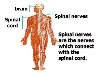
Overview of PNS Spinal Nerves. Guo Ling, MD, PhD Department of Anatomy. Overview of Nervous System. Central nervous System(CNS). Peripheral Nervous System(PNS). Spinal nerves. brain. CNS. spinal cord. NS. cranial nerves spinal nerves visceral nerves. PNS.

E N D
Overview of PNS Spinal Nerves Guo Ling, MD, PhD Department of Anatomy
Overview of Nervous System Central nervous System(CNS) Peripheral Nervous System(PNS) Spinal nerves brain CNS spinal cord NS cranial nerves spinal nerves visceral nerves PNS
Nerve fibers:They are long microstructures formed by neuronal processes which are enveloped by both thick/ thin myelin sheath and neurilemma; or are only covered by neurilemma. The fibers distribute in CNS or PNS. Nerve:It is a long & solid macrostructure into which numerous nerve fibers only in PNS are bundled.
Classification of PNS 1. Based on distributions of component: somatic nerve & viseral nerve 2.Based on functions of component: afferent(sensory) & efferent (motor) nerves 3. Based on combination of the above two items: (1) somatic nerve : sensory and motor nerves (2) viseral nerve: 1) sensory nerve 2) motor nerve (vegetative nerve / autonomic nerve) a. sympathetic nerve b. parasympathetic nerve 4. Based on relay stops of nervous informatiom (1) cranial ganglia as well as spinal ganlia (g) (2) sensary ganglia(8):spinal g, genulate g of CN7, cochlear g & vestibular g of CN8 ,super g & infer g of CN9, super g & infer g of CN10) (3) ganglia of vegetative nerve:1)ganglia of sympathetic nerve: paravetebral g (19~24) & prevertebral g (4):celiac g,aorticorenal g, super mesenteric g & infer mesenteric g) 2)ganglia of parasympathetic nerve:a.paraorganic ganglia in head (4): ciliary g of CN3, otic g of CN9, pterygopalatine g & submandibular g of CN7 b.paraorganic ganglia in body except those in head & intraorganic ganglia in body including those in head
Spinal Nerve (SN) Focus on the following items: 1.Buildup o f Spinal Nerve 2. Functional Features of Each Primary branch of SN 3.Composition and Position of Each Plexus of SN 4.Typical Nerves in Each Plexus (1)Innervation of skin areas (2)Innervation of muscular groups (3)Vulnerable places of nerve injury (4)Subsequent symptoms resulting from the Injury
Spinal Nerves 1.Formation:Each nerve consists of the ant.root and pos.root,which are conjoined together at an intervertebral foramen. 2.Division:31 pairs in total,8C(C 1- 8),12T(T1-12), 5L( L1-5), 5S( S1-5) & Co1. 61 separate nerves.
3. Branches of Each Spinal Nerves:4 pairs (1) meningeal branches (containiung sensory &sympatheticfibers-mixed nerves) (2) communicating branches(white& gray )→ sympathetic trunks (containing visceral motor fibers)(3)dorsal branches (mixed nerves) (4) anterior branches(mixed nerves,C,L,S&Co first form plexes ,and then send concrete nerves;while only T immediatedly gives off its own nerves.)
Focusing on the following items of anterior branch forming plexuses: 1.Compsition of each plexus 2.Position of each plexus 3.Representive nerves of each plexus (1) Invervation of specific skin areas (2) Invervation of concrete skeletal muscles (3) Places vulnerable to injury (4) Relative symptoms & signs
4.Cervical Plexus (1) Formation It is made up of the anterior branches from C1-4. (2) Location It lies in the upper part of the neck.
(3) Main Branches 1) Superficial branches(sensory) a. Greater auricular N b. Lesser occipital N c. Transverse N of neck d. Supraclavicular N The four are all sensory Ns and emerge from the middle point of the post border of the sternocleidomastoid,which is a fixed mark for anaesthenia pucture. 2) Deep branches(mixed N) e. phrenic nerve
(4) Distributions of Cervical Spinal Nerves 1) Greater auricular N 2) Lesser occipital N 3) Transverse N of neck 4) Supraclavicular N → The skin of neck The skin of sup. part of thoracic wall 5) Phrenic nerve → Diaphragm Diaphragmatic and mediastinal pleurae Pericardium ,liver, gallbladder, biliary system
5.Brachial Plexus (1) Formation: the ant. branches of C5~8 and most part of the ant. branch of T1 (2)Branch Transition: ant branches→sup,mid,inf trnuks →3 cords Lateral cord: a.musculocutaneous N →lat Antebrachial cutaneous N b.median N lat root Medial cord: c. median N. med.root d.ulnar N. Posterior cord: e.radial nerve f.axillary N. g.long thoracic N.→serratus ant. h.thoracodorsal N.→latissimus dorsi
(3). Location: lying in scalene fissure,supraclavicular fossa & axillary fossa thoracodorsal N. long thoracic N.
Axillary N innervates deltoid& the skin of shoulder
Skin of lat.side of forearm Lateral antebrachial cutaneous N
Radial N→ Triceps brachil Brachioradialis Extensor of the forearm ,the skin on radial side of dorsum of the hand,proximal digits of the lateral two & half fingers Radial N & its distribution
Ulnar N→ Flexor carpi ulnar Medial half of the flexor digitorum profoundus Ulnar half of the dorsum of the hand Post. Surface of the ulnar one and half fingers The muscles of hypothenar Interosseous muscles 3th, 4th lumbricales Adductor pollicis
Distributions of Median N Pronator Most of the flexor muscles except the brachoradialis the flexor carpi ulnaris Medial half of the flexor digitorum profoundus Thenar muscle except for adductor pollicis 1st,2nd lumbricales,the skin of thenar & central part of the palm, the skin of palmar aspect of the thumb, 2nd,3rd lateral half of 4th fingers & the dorsum of the middle & terminal phalanges
Humerus Fracture and Relaitve Injuries a. Fracture of surgical neck →axillary N →“winging of the scapula” b.Fracture of middle part of humerus →radial N →“wristdrop” c.Fracture of med epicondyle of humerus→ulnar N →“claw-like hand ” d.Fracture of lower end of humerus→median N →“ape-like hand ”
Innervation of Flexor and Extensor Muscles Acting on the Elbow Joint Flexing elbow joint:biceps brachii brachialis musculocutaneous N. brachioradialis Extending elbow joint:triceps brachii radial N.
Nerve Supply of Forearm Muscles a. brachioradialis ----radial N. b. ulnar half of flexor digitorum superficialis, flexor carpi ulnaris------------ulnar N. c. the remainingmuscles of anterior muscular group of forearm----median N. d. all muscles of posterior muscular group of forearm ---radial N.
Anterior Branches of Thoracic Nerves (Intercostal Nerves) Composition: 12 pairs 11 pairs of intercostal Ns. 1 pair of subcostal N Location:lying along the relative costal groove & going between the int﹠ext intercostal Ms Segmental arrangements: T2—-the sternal angle T4----the nipple T6----the xiphoid process T8----the costal arch T10----the umbilicus T12----the ant.superior iliac spine
Lumbar Plexus Composition: ant.branches of L1—3 & parts of the subcostal N and L4 Location: going within the psoas major ③ ④ Main branches: ⑤ ①Femoral N → → Saphenous N ① ② Obturator N ③ iliohypogastric N ④ilioinguinal N ⑤Lat. femoral cutaneous N ⑥Genitofemoral N ⑥ ②
Femoral N. Obturator N. Saphenous N.
Distributions of the Branches of Lumbar Plexus Femoral N---→muscles of anterior muscular group in the thigh (quadriceps femoris, sartorius, pectineus) Saphenous N →the skin of the medial side of the leg and foot Obturator N→muscles of medial muscular group in the thigh iliohypogastric N →the muscles of the lower part of the abdominal wall, the skin of hypogastric region & inguinal region ilioinguinal N → muscles of lower partof abdominal wall Lat. femoral cutaneous N →skin on ant.& lat.parts of the thigh Genitofemoral N →cremaster, scrotum(greater lip of pudendum)
Sacral Plexus Composition: the ant. branches of L5~8 ,the ant.branches of all the sacral Ns and coccygeal N. Location: in the pelvis, going on ant.surface of the piriformis, looking like a triangle shape.
Main Branches 1. sup.gluteal N. 2. inf.gluteal N. 3. post.femoral cutaneous N. 4. pudendal N. 5. sciatic N.
sup.gluteal N. inf.gluteal N. sciatic N. pudendal N. pos.femoral cutaneous N.
Pudendal N. →infrapiriformis foramina →the lesser sciatic foramen→the ischiorectal fossa,innervating the muscles and the skin of the perineum
Sciatic N Common Peroneal N Tibial N Superf.peroneal N deep peroneal N Medial plantar N lateral plantar N Sural N
Distributions of the Branches of Sacral Plexus: Sup.gluteal N→gluteal medius & minimus ,tensor faciae latae Inf.gluteal N→gluteal maxium & lower part of gluteal region Post.femoral cutaneous N→skin of the post part of the thigh Perineal N→the muscles of perineum & the skin of sacrum Sciatic nerve →muscles of post muscular group & skin in thigh Tibial N →muscles of post muscular group & skin of leg Med.& lat. Plantar N→plantar muscles & skin over the sole of the foot Sup.peroneal N→muscles of lat muscular group & skin of leg Deep peroneal N→muscles of ant muscular group & skin of leg Sural N→the skin of post.& lat. surfaces on the leg
Innervation of Muscles in Lower Limbs muscles of ant. group in thigh ---femoral N muscles of med. group in thigh ---obturator N muscles of post. group in thigh ----- sciatic N muscles of post. group in leg plantar muscles in foot-- -------------------tibial N muscles of lat. group in leg----- superficial peroneal N muscles of ant. group in leg------ deep peroneal N
Innervation of Muscles Acting on Knee Joint muscles nerves Movements Ms of post. group in thigh sciatic N. flexing knee joint(leg ) gastrocnemius tibial N. Extending knee joint(leg ) Quadriceps femoris femoral N.
Innervation of Skin of Foot saphenous N. →skin onmedial side of foot deep peroneal N. →dorsal skin between 1st &2nd toes sup. peroneal N. →most part of dorsum skin of foot tibial N. →sole skin of foot sural N. →dorsal skin on post.& lat.borders of foot