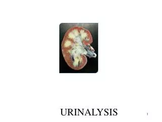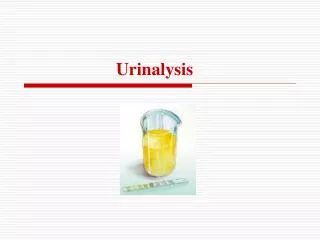
Urinalysis
Urinalysis. Prepared by Hamad ALAssaf alassaf_h@yahoo.com. Urine Examination. 1- Physical Examination 2- Chemical Examination 3- Microscopic Examination. URINE ANALYSIS Physical Examination. 1- Volume 2- Specific Gravity 3- Apperance 4- Color 5- Odor 6- Deposit


Urinalysis
E N D
Presentation Transcript
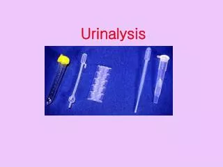
Urinalysis Prepared by HamadALAssaf alassaf_h@yahoo.com
Urine Examination 1- Physical Examination 2- Chemical Examination 3- Microscopic Examination
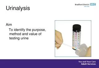
URINE ANALYSISPhysical Examination 1- Volume 2- Specific Gravity 3- Apperance 4- Color 5- Odor 6- Deposit 7- Reaction (pH)
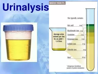
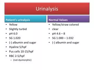
URINE ANALYSISPhysical Examination 1- Volume: Normal urine volume in 24 hours is 600-2000 ml A- Urine volume increases (Polyuria) in the following conditions: Physiological: • Increased fluid intake • Diuretic Pathological: • Diabetes mellitus • Diabetes insipidus • Chronic renal failure B- Urine volume decreases (Oliguria or anuria)in the following conditions: • Dehydration • Acute renal failure • Obstruction
URINE ANALYSISPhysical Examination 2- Specific gravity (SG): • Specific gravity measures solute concentration (urea and sodium). • Normallythe specific gravity ranges between 1.015-1.025. A- Increased in • Dehydration (with oliguria) • Diabetes Mellitus (with polyuria) • Acute renal failure (with oliguria) B- Decreased in • Diabetes insipidus (with polyuria)
URINE ANALYSISPhysical Examination 3- Appearance: - Normal fresh urine: clear (transparent) - Abnormal : Cloudy urine Indicates possible abnormal constituents such as: • White blood cells • Epithelial cells • Crystals • Bacteria N.B. Stored urine with no preservative & no cooling may turn clear urine samples into cloudyurine.
URINE ANALYSISPhysical Examination 4- Color: - Normal color: pale yellow (amber yellow) due to the presence of pigments of urobilinor urobilinogen - Abnormal colors of urine: • Colorless • Orange • Greenish yellow • Red • Black • Smoky
Left to right: (Straw, clear) (yellow, clear) (yellow, hazy) (yellow, clear) (red-orange, clear) (brown, hazy) Urine Colour
URINE ANALYSISPhysical Examination • Color(cont.) 1- Colorless Urine: • Chronic renal failure • Diabetes insipidus. 2- Orange Urine: • Ingestion of large amount of carotenoids (vitamin A) 3- Yellowish - brown urine: • due to presence of billirubin in cases of : • Obstructive Jaundice • Hepatic Jaundice
URINE ANALYSISPhysical Examination • Color(cont.) 4- Red urine: due to presence of blood,hemoglobin & RBCs. 5- Black urine: • Methemoglobin • Homogentisic acid in alkaptonuria • Malignant malaria (black water fever due to Malaria falciparum). • Melanin(melanoma) 6- Smoky urine: • presence RBCs. in the urine, in cases of acute glomerulonephritis
URINE ANALYSISPhysical Examination 5- Odor: • Normal Urineferous odor: The normal odor of fresh voided urine sample • Abnormal Odors: 1- Fruity odor: due to presence of acetone in the urine as in diabetic ketoacidosis 2- Ammonia odor: due to release of ammonia as result of: - the bacterial action on urea in the contaminated urine - or long standing exposed urine samples.
URINE ANALYSISPhysical Examination 6- Deposits: • Normally the urine is devoid of deposits. • The presence of deposits is mainly due to various types of crystals, salts and cells.
URINE ANALYSISPhysical Examination 7- Reaction (pH): • Normally: The pH of urine varies from 4.6 - 8.0 1- Acidic urine: • Large intake of meat & certain fruits (cranberries) • Metabolic & respiratory acidosis 2- Alkaline urine: • Vegetarians • Metabolic & respiratory alkalosis • Urinary tract infection by urea splitting bacteria which spliturea to ammonia (alkaline)
URINE ANALYSISChemical Examination • The presence of normal and abnormal chemical elements in the urine are detected using dry reagent strips. • These plastic strips contain absorbent pads with various chemical reagents for determining a specific substance. • When the test strip is put in urine the reagents are activated and a chemical reaction occurs. • The chemical reaction results in a specific color change.
URINE ANALYSISChemical Examination Used in the LAB for routine urine analysis (10 Chemical Tests) Used in Emergency Room (ER) for diagnosis of Diabetic Ketoacidosis (DKA) (3 chemical tests: Glucose, Ketones & protein)
URINE ANALYSISChemical Examination Abnormal Constituents of Urine 1- Proteins (proteinuria) 2- Sugars (glucosuria, fructosuria & galactosuria) 3- ketone Bodies (ketonuria) 4- Billirubin (billirubinuria) & Bile Salts 5- Nitrites
URINE ANALYSISChemical Examination 1- Proteins: (proteinuria) Proteinuria is divided into prerenal, renal and postrenalproteinuria. 1-Prerenal proteinuria: Bence-Jones protein: This abnormal gamma globulin (light chains only) is synthesized by malignant plasma cells (multiple myeloma). It precipitates at 60oC, redissolves at 100oC and reprecipitates on cooling. 2-Renal proteinuria: • Severe muscular exercise • After prolonged standing • Acute glomerulonephritis • Nephrotic syndrome 3- Postrenal proteinuria: • Lower urinary tract inflammation, tumors or stones.
URINE ANALYSISChemical Examination 2- Glucose: (glycosuria) Presence of detectable amount of glucose in urine which occurs in the following conditions: • Uncontrolled Diabetes Mellitus (DM) • Renal glucosuria with lowering of renal threshold : e.g. during pregnancy (gestational diabetes). - Fructose(Fructosuria) Presence of fructose in urine & may be due to: - Alimentary causes following the ingestion of large amounts of fructose - Fructosemia & hereditary fructose intolerance (Metabolic disorders of fructose). - Galactose(Galactosuria): Presence of galactose in urine& may be due to: - Alimentary causes following the ingestion of large amount of galactose. - Galactossemia
URINE ANALYSISChemical Examination 3- Ketone Bodies (Ketonuria): Presence of acetone, acetoacetic acid & βhydroxybutyricacid in urine due to: • Diabetic ketoacidosis(uncontrolled DM) • Starvation • Unbalanced diet: high fat & low carbohydrates diet.
URINE ANALYSISChemical Examination 4- Bilirubin (bilirubinuria) • Billirubin appears in urine in cases of: • Hepatocellular Jaundice: as in viral hepatitis • Obstructive Jaundice as any cause of obstruction of bile duct 5- Nitrites: • In bacteruria in urine (in cases of Urinary Tract Infection, UTI)
URINE ANALYSISMicroscopic Examination • The urine specimen is centrifuged and the liquid portion is poured off.
URINE ANALYSISMicroscopic Examination • The concentrated cellular sediment, is then placed on a microscope slide, covered with a coverslip and read under microscope.
URINE ANALYSISMicroscopic Examination A variety of normal and abnormal cellular elements may be seen in urine sediment such as: • Casts • Crystals • Amorphus • Microorganisms • Pus cells • RBCs • Epithelial cells • Mucus
URINE ANALYSISMicroscopic Examination RBCs 40x objective - presence of a few is normal (2 – 5 cells/HPF) - higher numbers are indicator of renal disease - result of bleeding at any point in urinary system
URINE ANALYSISMicroscopic Examination Pus cells 40x objective • a few are normal (2 – 4 cells /HPF) • high numbers indicate inflammation or infection somewhere along the urinary or genital tract.
URINE ANALYSISMicroscopic Examination Mucus 40x objective • look like long, ribbon-like threads • common finding in urine sediment • secreted by glands in the lower urinary tract. • Become more with UTI, ulcerative colitis, kidney stones
URINE ANALYSISMicroscopic Examination Epithelial Cells 40x objective • cells are large and flat • normal cells that line the urinary and genital tract or renal tubules
URINE ANALYSISMicroscopic Examination Crystals of calcium oxalate colorless octahedron found in acid urine Crystals of triple phosphate colorless, “coffin-lid” prism common finding; no clinically significant
URINE ANALYSISMicroscopic Examination Amorphous urate (Acidic urine) Amorphous phosphate (alkaline urine)
URINE ANALYSISMicroscopic Examination Bacteria Yeast
URINE ANALYSISMicroscopic Examination (Parasites) schistosomahaematobium Trichomonasvaginalis Characterized by Central spin It is not Urinary system protozoa, it’s vagina protozoa
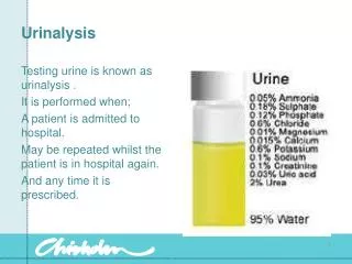
It’s not just water ! Thank you