URINALYSIS
URINALYSIS . Clinical Textbook for Veterinary Technicians 4 th Ed. Dennis M McCurnin Saunders. Urinalysis is a clinical laboratory procedure that should be performed as part of any minimum data base . However, it is often skipped. Tests should be run on fresh urine when possible.

URINALYSIS
E N D
Presentation Transcript
URINALYSIS Clinical Textbook for Veterinary Technicians 4th Ed. Dennis M McCurnin Saunders
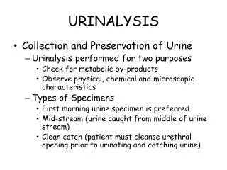
Urinalysis is a clinical laboratory procedure that should be performed as part of any minimum data base . However, it is often skipped. Tests should be run on fresh urine when possible. Urine samples should be collected into clean glass or plastic containers – in general no preservatives are necessary. The best time to collect urine is usually first thing in the morning - urine that accumulates during the relative inactivity of the night is less likely to be influenced by feeding or exercise. Morning urine is concentrated and therefore, if they are present, contains the abnormal constituents.
URINE COLLECTION • Free-Flow – the simplest method, if the animal cooperates • the initial stream should be avoided – it may contain cells and debris from the • urethra and lower genital tract. • it is best to catch a mid-stream sample. • Cystocentesis- this procedure involves placing a needle with a syringe attached • through the ventral abdominal wall into the lumen of the bladder and aspirating • the urine. • aseptic technique must be used • iatrogenic hemorrhage may occur • iatros = healing or physician • gennan = as a product of • iatarogenic = due to the doctor • Catheterization – collecting urine by catheterization of the bladder • must be done as aseptically as possible to prevent introduction of bacteria into • urinary tract • care should be taken to avoid traumatizing the lining of the urethra
Cystocentesis Catheterization Cyst = sac containing fluid Centesis = surgical puncture Cystocentesis = surgical puncture of the urinary bladder
No matter how the urine is collected, it should be analyzed as soon as possible. Many changes begin to occur in urine almost immediately. * bacteria will multiply * cells may degenerate * casts may dissolve * bacteria may produce urease that will convert urea to ammonia causing pH to increase If there is to be a delay in analyzing the sample, it should be refrigerated. PHYSICAL PROPERTIES Color - Yellow or Amber = normal Bright red = hematuria = RBCs in the urine Reddish Brown = hemoglobinuria or myoglobinuria = hemoglobin in the urine yellow-brown= if forms yellow foam when shaken = may have high concentrations of bilirubin or urobilin
Normal Urine Hemoglobinuria Hematuria
Turbidity = cloudy ( fresh urine is usually transparent) However as urine cools, some salts may precipitate, causing the urine to become cloudy. Fresh urine that is cloudy (except in equine urine) is often pathological. It must be examined microscopically to determine the cause: * pus * mucus * bacteria * casts * crystals Note: Fresh equine urine is normally cloudy because it contains mucus and calcium carbonate crystals
Specific Gravity (SG) – one of the most important parts of a urinalysis Must be determined before urine has been centrifuged. The SG value depends on the number and size of the particles in solution and is an indicator of the ability of the kidney to concentrate urine. The SG is determined with a refractometer.
Chemical Evaluation Reagent strip chemistries should be performed on unspun urine, unless the urine is very turbid. If the sample is turbid, the test should be done after the sample has been centrifuged. Protein, glucose, ketones, blood and bilirubin in the urine are routinely determined as well as urinary pH.
pH pH is used to express the hydrogen ion concentration . Readings above 7 are said to be alkalotic. Readings below 7 are said to be acidotic. In general, dogs and cats tend to have more acid urine than do cattle and horses.
Protein Protein loss into the urine can be a significant drain on the body’s protein stores. In interpreting the cause for positive urine protein test, one must also consider the results of the test for blood and the microscopic examination of sediment. There are few external factors that may temporarily alter glomerular permeability of protein and result in proteinuria – shock, severe exercise, cardiac or central nervous system disease, and the postcolostral period in neonates.
When excessive hemorrhage into the urinary tract occurs the test for protein will be positive and erythrocytes will be seen in the sediment. Possible causes for urinary tract hemorrhage include trauma, neoplasia, and inflammation. When proteinuria occurs in conjunction with increased leukocytes in the urine sediment inflammation of the urinary tract should be suspected. In some urinary tract infections, bacteria may be seen in the sediment.
Glucose Another common reagent strip test for urine is the test for glucose. Normally, urine contains no detectable glucose. Glucose is filtered by the glomerulus, but the body preserves this energy source by reabsorbing it in the proximal renal tubules. The primary cause for glucosuria, therefore, is hyperglycemia. Diabetes mellitus is a common cause of hyperglycemia and glucosuria.
Ketones Ketones will appear in the urine before they build up to a detectable level in the bloodstream, so ketonuria may occur before a detectable ketonemia occurs . Ketonuria indicates excessive fat metabolism, a deficiency in carbohydrate metabolism, or both, but is most commonly seen in conjunction with glucosurea as a complication of diabetes mellitus.
Bilirubin There are reagent strips and tablets for detecting bilirubinuria. Bilirubin in urine can also be crudely detected by the foam test. If a yellowish foam appears when the urine sample is shaken, bilirubin is likely present. Many normal dogs, and sometimes cattle, will exhibit bilirubinuria because the kidneys , as well as the liver, have an enzyme that can conjugate bilirubin. This, however, is lacking in cats, and any bilirubinuria is a significant abnormality in cats.
Blood The designation “blood” is somewhat misleading because this test actually detects intact RBCs, hemoglobin, and myoglobin. If the urine is red and cloudy and erythrocytes are present in the sediment, hematuria is the reason for the positive blood reaction. Hemoglobinuria results in red to brown urine with a positive urine blood reaction and no erythrocytes in the sediment. A positive urine protein test may also be apparent. In contrast to hematuria, hemoglobinuria will be accompanied by hemoglobinemia, imparting a pink to reddish discoloration to the serum or plasma.
Myoglobinuria will similarly result in red to brown urine with no erythrocytes in the sediment, a positive urine blood reaction, and a positive urine protein test. However, during myoglobinuria, the blood serum or plasma generally remains clear. Animals with myoglobinuria generally do not show evidence of anemia but have some type of muscle disease such as exertionalmyopathy in horses, trauma, electrical shock, or pressure necrosis from prolonged recumbencey.
Urine sediment examination may reveal extremely useful diagnostic information and be crucial for correct interpretation of the chemical analyses. A few cells and a few casts may be found in normal urine, but increased numbers of various elements indicate certain diseases. Epithelial Cells Three types of epithelial cells can be found in urine sediment: Squamous: large, with angular borders and small nuclei originating from the lining of the distal urethra and vagina or prepuce and not generally indicative of diseases. Transitional : medium sized and oval, spindled, or caudate cells found lining the proximal urethra, bladder ureters, and renal pelvis Renal tubular : small, round cells - may be indicative of tubular degeneration
Blood Cells or Erythrocytes in unstained sediment appear colorless or yellowish and are round and slightly refractile, with no internal structure. They may be confused with fat droplets, but erythrocytes are fairly uniform in size and do not float in and out of planes of focus as do fat droplets. If there is doubt, a drop of diluted acetic acid will lyse erythrocytes helping to differentiate them from fat droplets. In concentrated urine, erythrocytes may lose fluid and become creatnated. In dilute urine, they may imbibe water and swell or even lyse, becoming ghost cells. Leukocyte or White blood cells in urine sediment are round and granular and larger than erythrocytes but small than epithelial cells. The presence of more than five to eight WBCs per high powered field indicates inflammation of the urinary or urogenital tract depending on the method of collection. When this occurs, a careful check for bacteria should be made.
CASTS Casts are another prominent feature of urine sediment. They are elongated structures composed of protein from plasma and mucoprotein from the renal tubules. In general , they form in the distal tubules, in which the urine is more concentrated and acidic. Any structures that happen to be in the tubules at the time the casts form (erythrocytes, leukocytes or epithelial cells) become embedded in the casts. The presence of increased numbers of casts helps to localize the renal disease to the tubules, but the numbers do not necessarily correlate with the severity of disease.
(1) Hyaline casts are colorless, homogeneous, and semitransparent. They may be difficult to see unless the light is reduced. They indicate mild glomerular leakage. Cellular casts contain recognizable cells imbedded in the protein matrix. They may be epithelial cell casts that contain sloughed tubular or leukocyte casts that indicate renal inflammation or pyelonephritis. Granular casts are derived from degenerating cells or cellular casts. They are characterized by a nonspecific granular matrix and are designated either coarsely or finely granular depending on the degree of degeneration. They are probably the most common type of casts found in animals. Waxy casts are wide and homogeneous, usually with distinct blunt or squared ends. They are the next stage of degeneration from granular casts and indicate a more chronic tubular lesion.
Fatty casts contain fat globules from degenerating tubular epithelial cells and are most common in cats because of the high lipid content of feline tubular epithelium. Crystals Crystal precipitation and presence depend on urine pH and the solubility and concentration of the substance forming them. Crystals are common and most are not indicative of disease. In carnivores, phosphates, urates and oxalates are normal, whereas in herbivores, calcium carbonate crystals are the most common. Urine crystals that accompany pathological conditions include ammonium biurate, Monohydrate calcium oxalate, bilirubin and cystine crystals.
Microorganisms Bacteria in unstained urine sediment may be difficult to detect. Rods may appear singly or in chains, but cocci may be lost in brownian movement. For this reason, whenever bacteria are suspected, the sediment should be stained to examine it more thoroughly. Bacteria in a voided sample often are not significant because they may be normal flora from the distal urogential tract, especially from the prepuce or vagina. Bacteria are significant if they occur in catheterized or cystocentesis samples. Their presence should be correlated with leukocytes in urine because bacteria with no leukocyte response should raise suspicion of contamination of the sample. If bacteria are present in a urine sample, they can multiply as time passes, so this should be taken into consideration when the sample is being analyzed.
Rarely, fungal organisms are found in urine sediment. The majority of these are insignificant contaminants, although some such as Blastomyces, may be significant. Miscellaneous Usually insignificant components of urine sediment include mucus, fat, sperm and parasites. Mucus appears as narrow, twisted, robbon like strands. It is normal in equine urine and can be seen in other species due to genital secretions or irritation of the urethra. Fat as previously noted, takes the form of refractile, variably sized spheres in many planes of focus. Fat is rarely significant. Sperm are commonly seen in male canine urine. Parasites that can occur in urine include the ova of Stephanurusdentatus, Dictophymarenale, and Capillariaplica and microflilaria of Dirofliariaimmitis.
Squamous epithelial cells transitional epithelial cells
Hyaline cast Cellular cast Granular cast Waxy Cast
Fatty Cast Phosphate crystals Urate crystals Oxalate crystals
Calcium carbonate crystals Ammonium biurate crystals Bilirubin crystals Monohydrate calcium crystals
Cystine crystals Bacteria and yeast