Urinalysis
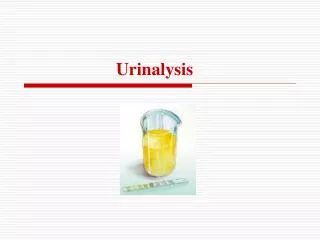
Urinalysis. Routine Urinalysis 1) 물리적 검사 - color, turbidity & odor - specific gravity ( 비중 ) 2) 화학적 검사 3) microscopic examination - pH - RBC & WBC

Urinalysis
E N D
Presentation Transcript
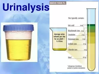
Routine Urinalysis • 1) 물리적 검사 • - color, turbidity & odor • - specific gravity (비중) • 2) 화학적 검사 3) microscopic examination • - pH - RBC & WBC • - protein - epithelial cells • - glucose - casts • - ketone - crystals • - bilirubin - microorganisms • - urobilinogen • - occult blood • - nitrite • - leukocyte
2. Special tests 1) 물리적 검사 - 12 or 24 hr urine volume - osmolarity 2) 화학적 검사 - protein : Bence-Jones protein - specific amino acid or metabolic product : alkaptouria - carbohydrate : lactose, fructose, galactose - hemosiderin - porphyrins : uroporphyrin, coproporphyrin - melanin - indican - hormone · 5-HIAA (hydroxy indole acetic acid) : serotonine metabolite test · 17-OHCS (hydroxycorticosteroids) · 17-KS (ketosteroids) · VMA (vanillylmandelic acid) · HCG (human chorionic gonadotropin) 3) 현미경 검사 - Addis count (뇨침사 정량, 12시간뇨 사용; WBC, RBC, EP cells, Cast 정량)
1-1) 물리적 검사 1. Color 정상 straw (일반 정상뇨), yellow (일반적으로 뇨량 적고, 음식 또는 약물 영향) amber (요량 적고 비중 높은 농축뇨), colorless (요량 많고 비중 낮은 희석뇨) 비정상 무색 : 당뇨, 뇨붕증, 다뇨 적색 : hematuria, hemoglobinuria, myoglobinuria, porphyrinuria 오렌지색 : bile pigment, carotene, food, 농축뇨 녹색~청록색 : bile pigment, vitamin B complex, dye(methylene blue) 흑색~암갈색 : alkaptouria, melanin, bilirubin 유백색 : filaria에 의한 chyluria(유미뇨), fat, pus, 인산염류 2. Turbidity 정상 투명 : 무정형 인산염 : 중성 또는 alkali 뇨에서 백색 침전, 5~10% acetic acid 첨가시 투명화 무정형 뇨산염 : 뇨의 온도가 내려가고 산성뇨에서 침전, 가열 또는 NaOH 가하면 용해 비정상 농 (pyuria), 혈액, 상피세포가 많으면, 혼탁 세균뇨, lipiduria, filaria에 의한 chyluria
Turbid urines Colored urines
1-2) 화학적 검사 1. pH (5.5~6.5) 복합 pH 지시약 (bromthymol blue + methyl red), acid : orange, alkali : blue 2. 비중 (수시뇨 : 1.003~1.030, 24시간뇨 : 1.015~1.025) 뇨량 많으면 비중 낮고, 뇨량 적으면 비중 높다.(ex, 요붕증 : 뇨량 많고 비중 낮음) (예외, 당뇨 : 뇨량 많고 비중 높음) 측정법 Multistix SG 10 : electrolyte (Na+, K+ in sample) + COOH H+방출 H+ + Bromthymol blue color change 굴절계 : 뇨비중, serum or plasma protein 요비중계 : 3. 단백뇨 요단백의 원인 a) 1일 protein 배설량 150mg 이상 일 때 (사구체 이상으로 혈청 단백 누출) b) 세뇨관에서의 protein 재흡수 장애 c) Bence-Jones protein 측정법 a) strip : tetrabromphenol blue(TBPB)을 지시약으로 사용 (혈액, 농, 질분비물 등에 의해 위양성, pH 9.0이상 알카리에서 위양성 가능성) b) Sulfosalicylic acid test (20%) : 단백과 침전 민감도 : strip (15~20mg/dl), SSA (5mg/dl, 민감도 가장 우수)) c) Trichloroacetic acid (TCA) : turbidity로 24시간 urine protein 측정 d) Esbach법 (picric acid, citric acid) e) Tsuchiya법 (phosphotungstic acid)
굴절계 Urine strip SSA for urine protein Clinitest for urine sugar
Bence-Jones protein : 40~50ºC (혼탁), 60~70ºC (침전), 85- 100ºC (용해) α2-globulin 혹은 β-globulin 분획 위치에서 peak 다발성 골수종의 진단 4. 당뇨 (urine sugars) 신장의 신역치 : 160~180mg.dl 검사법 a) 포도당 산화효소 시험지법 GOD β-D-glucose + O2 gluconic acid + H2O2 POD H2O2 + chromogen H2O + oxidized chromogen * ascorbic acid(50mg/dl), ketone body(40mg/dl) 존재시 위음성 b) Benedict 법 heat CuSO4 + glucose Cu2O + oxidized glucose (blue) alkali (red) * sucrose를 제외한 모든 환원당 또는 비환원당과 반응 * 각종 환원성 당의 감별검사법 lactose : Malfatti 법, fructose : Seliwanoff 법 pentose : Bial orcinol 법, galactose : Mucic 법
요당 검사법에 따른 반응 관계 성분 GOD (시험지법) Cu 환원법 Glucose 양성 양성 Fructose 영향 없음 양성 Galactose 영향 없음 양성 Lactose 영향 없음 양성 Maltose 영향 없음 양성 Pentose 영향 없음 양성 Sucrose 영향 없음 영향 없음 @ Benedict test 3분간 boiling benedict soln. Benedict soln. + glucose
5. Ketone body(acetone, acetoacetic acid, β-hydroxybutyric acid를 총칭) 지방대사의 분해 산물 시험지법에서는 acetone, acetoacetic acid 2가지만 반응하고 β-hydroxybutyric acid와는 반응하지 않는다. 당뇨, 기아 등 glucose 이용이 저하되면 체내 ketone 축적 ---뇨로 배설(ketouria) 당뇨병성 ketoacidosis의 진단에 유용 검사법 a) 시험지법 Lange 반응 acetic acid, acetone + Na-nitroprusside 자홍색 (적자색) alkali b) Hart-Lange test (β-hydroxybutyric acid 측정법) 6. Occult blood 미량 적혈구, Hb 측정법 혈뇨, 혈색소뇨, 미오글로빈뇨의 차이점 Hematuria Hemoglobinuria Myoglobinuria Microscopic POS NEG NEG Chemical POS POS POS 검사법 a) 시험지법 Blood + H2O2 + chromogen H2O 산화형 chromogen POD activity * chromogen : tetramethy benzidine (녹색~암청색), O-toluidine (청녹색)
Occult Blood (chromogen : O-toluidine) POSITIVE NEGATIVE b) Benzidine test c) O-toluidine test d) Guaiac test @ Myoglobin 측정법 (urine color가 red - brown 일때 실시) 80% ammonium sulfate : Hb과 다른 단백질 침전, Mb 침전 못시킴 (상층) sangur test – 상층액 (pos), 침사액 ( neg) (확인 검사 필요) 포화 ammonium sulfate : Mb 침전 sangur test – 상층액 (neg), 침사액 ( pos) (Mb 양성판정)
@ Hemosiderin 측정법 혈색소뇨의 원인이 되는 급성 용혈성 질환이 있은지 2~3일 후에 요의 침사에 출현 검사법 : prussian blue 7. Bilirubin 비리루빈 origin : Hb (75%), Mb (15%), catalase, cytochrome (10%) • 용혈성 황달 : 용혈성 빈혈의 원인 • 소변 : urobilinogen (++), bilirubin (-), • 분변 : uribilinogen (++++) • 혈청 : 간접 bilirubin 증가 • 간세포성 황달 : 간염 등 간세포 손상이 원인 • 소변 : urobilinogen (+++), bilirubin (++++) • 분변 : urobilinogen (+, 감소경향) • 혈청 : 직접 bilirubin 증가 • 폐쇄성 황달 : 담관 폐쇄가 원인 • 소변 : urobilinogen (-), bilirubin (++++) • 분변 : urobilinogen (-, 감소) • 혈청 : 직접 bilirubin 증가
Bilirubin 검사법 a) 시험지법 coupling reaction Bilirubin + diazo reagent azobilirubin 강산성 * diazo reagent = 2-methyl-5-nitroaniline + sodium nitrite b) Gmelin test bilirubin을 산화(reagent, yellow nitric acid)시켜 경계면에 녹색의 biliverdin이 형성 c) Icto test bilirubin이 디아조와 반응하여 적보라색으로 변화 bilirubin 검출법 중 민감도 가장 우수 8. Urobilinogen bilirubin 대사 종산물 환원 산화 conjugated bilirubin urobilinogen Urobilin (뇨) / stercobilin(분변) 장내세균 검사법 a) 시험지법 : urobilinogen + p-methoxybenzole diazonium fluoborate 적색의 carmine 강산 b) Erhlich aldehyde 법 : p-dimethylaminobenzaldehyde와 반응하여 적색 정상치 : neg ~ trace
9. Nitrite 신장, 비뇨기계의 셰균감염 예비진단 nitrate(질산염)가 세균에 의해 nitrite(아질산염)으로 환원 검사법 nitrite + arsanilic acid diazonium 화합물 coupling reaction diazonium 화합물 + tetrahydrobenzoquinoline pink azo 색소 10. Leukocyte esterase 호중구의 esterase 이용 발색 유도 고배율 현미경에서 5개 이상 존재하면 양성 반응 뇨로의 감염 여부를 알아보는 선별검사 11. Ascorbic acid Vit. C의 농도가 25mg/dl 이상 높게 나오면 요당, occult blood, bilirubin, leukocyte, nitrite 검사에 영향 줌. (반응저해)
1. 뇨의 채취 일반 뇨검사 수시뇨(random urine) 아침 첫뇨(first morning urine) 정량검사 24시간 뇨 (화학 검사용) 12시간 뇨 (Addis count : WBC, RBC, EP cell, cast 정량 계산) 세균 배양 : 중간뇨 채취 세포검사 : 세포검사 (50% alcohol 과 뇨 동량 혼합) 2. 정상뇨량 24시간 뇨량 : 600~2,000ml(성인) 주간뇨와 야간뇨의 비율은 3:1 ~ 4:1 3. 뇨 보존법 냉장 : pH, 세포 성분의 변화, 세균의 성장 발육 억제 냉동 : 화학분석 위한 장기간 보존 pH 조절 : pH 1~2 (catecholamine, VMA, 5-HIAA) pH 3 (amino acid)
화학보존제 a) Toluene : 화학적 성분 보존에 가장 좋은 보존제 (공기와 접촉 막아 산화 방지) b) Formalin : 세포성분 보존에 가장 좋은 보존제 (indican의 Obermyer 시험 방해, occult blood 방해, alkali copper 이용한 반응에서 환원작용하여 요당검사 위양성) c) Thymol : d) Conc HCl : VMA, catecholamine, 5-HIAA 검사에 사용 e) Glacial acetic acid : 5-HIAA f) Sodium fluoride(NaF) : glycolysis 억제 요의 실온 방치에 의한 성분 변화 item change item change color 진한 황색 혼탁도 증가 냄새 암모니아 냄새 pH alkali화 glucose 감소 urobilinogen 감소 bilirubin 감소 ketone 감소 occult blood 초기 용혈로 증가하나 후에는 POD 활성저하로 둔화 nitrite 약간 증가 뇨침사 관찰 곤란
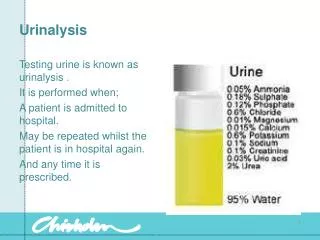
뇨의 참고치 pH : 5.5 ~ 6.5 (5.0 ~ 8.0) 비중 : 1.003 ~ 1.030 Glucose : 0 ~ 30 mg/dL ( 30 ~ 50 mg/dL = ; 70mg/dL = +1 ) Protein : 0 ~ 20 mg/dL ( 30 ~ 70 mg/dL = +1 ) Ketone body : Not detected Occult blood : Not detected Bilirubin : Not detected Urobilinogen : = 0.2 ~ 1.0 mg/dL Nitrite : not detected 뇨의 구성 성분 Glucose : < 300mg/L Protein : < 150mg/L /24hr Urea nitrogen : 7 ~ 16g/L/24hr Uric acid : 300 ~ 800 mg/24hr Creatinine : 1.2 ~ 1.8g/24hr
4. 뇨침사 검경 채뇨 1시간 이내의 신선뇨로 검경 뇨침사 성분의 origin 신전성 crystal 지방 신 유래 신성 cast 소원형 상피세포 신후성 적혈구, 백혈구 요로 유래 편평상피세포 정자 외래성(contamination) 세균 진균, 원충 이물질, 조영제 뇨침사 원심속도 : 1500rpm (5분), 2000rpm(3분) 세균뇨 원심속도 : 3000rpm (10분) 요 침 사
뇨침사의 종류 1) 유기침사 a) RBC b) WBC c) Epithelial cell (상피세포) - 소원형세포 (small round cell) - 이형상피세포 (transitional epithelial cell) - 편평상피세포 (squamous epithelial cell) d) Cast (원주체) - hyaline cast (초자원주) - granular cast (과립원주) - waxy cast (납양원주) - RBC cast (혈구원주) - WBC csat (백혈구 원주) - fatty cast (지방원주) - epithelial cast (상피원주) - cylindroid (유원주) e) Microorganism - bacteria - Trichomonas vaginalis - yeast like cell (candida) - parasite f) Spermatozoa 2) 무기침사 a) Amorphous material - acid urine : amorphous urate - alkali urine : amorphous phosphate b) Crystal <정상 산성뇨 결정> - calcium oxalate (수산칼슘) - uric acid (뇨산) - sodium urate (뇨산 나트륨) - calcium sulfate (황산칼슘) <비정상 산성뇨 결정> - leucine - tyrosine - cystine - bilirubin - hippuric acid - cholesterol - sulfonamide - hemosiderin <정상 알카리뇨에서 나타나는 결정> - triple phosphate - ammonium urate - calcium phosphate - calcium carbonate - amorphous phosphate
정상인 뇨침사 참고치 RBC : 0~2 RBC/HPF WBC : 0~2 WBC/HPF Cast : not found, only hyaline cast/LPF
2. Special Tests @ Indican 아미노산인 tryptophan이 부패되어 생성 검사법 a) Jaffe법 b) Obermayer법 @ Porphyrin Heme의 전구물질 중금속 중독의 지표 물질 Porphyrin 합성과정 glycine + succinyl CoA δ-aminolevulinic acid porphobilinogen urophophyrin coproporphyrin protoporphyrin + Fe Heme 검사법 a) Porphobilinogen 정성검사 (watson-Schwartz test) Porphobilinogen는 Ehrlich의 p-dimethylaminobenzaldehyde reagent와 반응하여 적색 발현 (유기용매의 용해도에 따라urobilinogen이나 indole의 적색은 chloroform 에 추출 용해되고 porphobilinogen은 용해되지 않음) 판정 : 양성—상층이 적색 또는 짙은 pink , 하층은 무색 또는 엷은 노랑, 갈색 (아래층,chloroform층이 적색이면 urobilinogen이나 indole의 존재 의미) b) Porphyrin 정성검사 porphyrin을 ether로 추출하여 적색 형광 검출
@ Phenylketonuria Phenylketonuria는 phenylalanine 대사의 선천적 장애로 발생 phenylalanine hydroxylase의 결핍으로 phenylalanine을 tyrosine으로 전환 시킬 수 없기 때문에 phenylpyruvic acid, phenyllactic acid, phenylacetic acid, o-oxyphenyl acetic acid가 뇨중에 다량 존재하게 되고 이를 총칭하여 phenylketouria라고 한다. * phenylketonuria, tyrosinosis, alkaptouria 대사 맟 장애 phenylalanine Phenylalanine hydroxylase blocked in phenylketonuria tyrosine p-hydroxy phenylpyruvic acid p-hydroxy phenylpyruvic acid oxidase blocked in tyrosinosis homogentisic acid homogentisic acid oxidase blocked in alkaptouria maleylacetoacetic acid 검사법 : Guthri test (special phenylalanine dependent strain B. subtilis 이용) 혈액에서의 phenylalanine 대사이상 검사법
@ Alkaptouria 유전성 대사이상으로 homogentisic acid oxidase의 결핍으로 발생 뇨의 배뇨시 정상으로 보이나 공기 중에 방치하면 갈색 또는 흑변함 @ Melanine Tyrosine의 종산물로 방치해 두면 흑색뇨로 변함 @ Urine Ca 검사법 : Sulkowitch test (ammonium oxalate)를 이용하여 침전시킨 후 혼탁 측정 @ Vanyllylmandelic acid (VMA) catecholamine은 epinephrine, noepinephrine, dopamine으로 구성 VMA는 epinephrine, noepinephrine의 최종 대사산물로 catecholamine 생산량 반영 함 @ 17-OHCS glucocorticoids의 분비 이상이 의심되는 부신피질 질환에서 검사 측정법 17-OHCS-glucuronide 결합을 β-glucuronidase를 이용하여 17-OHCS를 가수분해 Porter-silver 반응(phenylhydrazine과 17-OHCS 반응) 이용하여 황적색 측정 @ 5-HIAA serotonine의 주요 대사산물 @ 17-KS Zimmermann 반응 이용하여 androgen의 분비량 측정
@ Osmolarity 뇨의 삼투압 측정 (수분이나 전해질 대사의 좋은 지표, 신장 세뇨관의 기능 파악) 측정원리 : 용매에 용질을 가하면 용매분자의 chemical potential이 용질분자의 mole에 비례하여 낮아지며 이로 인해 증기압이나 빙점, 삼투압의 증가, 비점의 증가가 나타난다. 측정방식 : 빙점강하, 증기압 증가 체액검사 위액의 화학적 검사 : 1) Congo red : pH 3.5 ~ 5.0에서 반응 2) Topffer 시약 : pH 2.8 ~ 4.2에서 유리염산과 반응 등황색 황색 수액의 globulin 정량 : 1) Nonne-Apelt 반응 2) Pandy 반응 CSF에서 가장 많이 출현하는 분획 : albumin 분변의 갈색 색소 : stercobilin
URINE SEDIMENTS Red blood cells abnormal RBC's white blood cells Oval fat bodies
Amorphous urates (Na, K, Mg, or Ca salts) Tyrosine crystals Capillaria plica is a helminth parasite Microfilariae of Dirofilaria immitis