URINALYSIS
URINALYSIS. The urinalysis is a fundamental test that should be performed in all urologic patients A complete urinalysis includes both chemical and microscopic analyses. . Reasons for inadequate urinalyses include. Improper collection, Failure to examine the specimen immediately,

URINALYSIS
E N D
Presentation Transcript
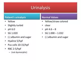
The urinalysis is a fundamental test that should be performed in all urologic patients • A complete urinalysis includes both chemical and microscopic analyses.
Reasons for inadequate urinalyses include • Improper collection, • Failure to examine the specimen immediately, • Incomplete examination (eg, most laboratories do not perform a microscopic analysis unless it is specifically requested by the provider), • Inexperience of the examiner, and (5) Inadequate appreciation of the significance of the findings.
Collection of Urinary Specimens • In the male patient, a midstream urine sample is obtained • The four aliquots have been designated Voided Bladder 1, Voided Bladder 2, Expressed Prostatic Secretions, and Voided Bladder 3 (VB1, VB2, EPS, and VB3) • To evaluate for a possible infection in a female, a catheterized urine sample should always be obtained. • All urine samples should be examined within 1 hour of collection and plated for culture and sensitivity if indicated for Neonates and Infants
Physical Examination of Urine • The physical examination of the urine includes an evaluation of color, turbidity, specific gravity and osmolality, and pH.
Color • The normal pale yellow color of urine is due to the presence of the pigment urochrome • Urine color varies most commonly because of concentration, but many foods, medications, metabolic products, and infection may produce abnormal urine color.
Turbidity • Cloudy urine is most commonly due to phosphaturia • The large numbers of white blood cells cause the urine to become turbid. • Pyuria is readily distinguished from phosphaturia either by smelling the urine (infected urine has a characteristic pungent odor) • Rare causes of cloudy urine include chyluria (in which there is an abnormal communication between the lymphatic system and the urinary tract resulting in lymph fluid being mixed with urine), lipiduria, hyperoxaluria, and hyperuricosuria.
Specific Gravity and Osmolality • Specific gravity of urine is easily determined from a urinary dipstick and usually varies from 1.001 to 1.035. • A specific gravity less than 1.008 is regarded as dilute, and a specific gravity greater than 1.020 is considered concentrated • Conditions that decrease specific gravity include -- • increased fluid intake, (2) diuretics, (3) decreased renal concentrating ability, and (4) diabetes insipidus. • Conditions that increase specific gravity include-- (1) decreased fluid intake; (2) dehydration owing to fever, sweating, vomiting, and diarrhea; (3) diabetes mellitus (glucosuria); and (4) inappropriate secretion of antidiuretic hormone. • Osmolality is a measure of the amount of material dissolved in the urine and usually varies between 50 and 1200 mOsm/L.
pH • Urinary pH is measured with a dipstick test strip( methyl red and bromothymol blue), which yield clearly distinguishable colors over the pH range from 5 to 9. • Urinary pH may vary from 4.5 to 8; • The average pH varies between 5.5 and 6.5. • A urinary pH between 4.5 and 5.5 is considered acidic, whereas a pH between 6.5 and 8 is considered alkaline.
In patients with a presumed UTI, an alkaline urine with a pH greater than 7.5 suggests infection with a urea-splitting organism, most commonly Proteus. • Urinary pH is usually acidic in patients with uric acid and cystine lithiasis. • Alkalinization of the urine is an important feature of therapy in both of these conditions
Chemical Examination of Urine • Urine dipsticks provide a quick and inexpensive method for detecting abnormal substances within the urine • The abnormal substances commonly tested for with a dipstick include • blood, (2) protein, (3) glucose, (4) ketones, (5) urobilinogen and bilirubin, and (6) white blood cells.
Hematuria • Normal urine should contain less than three red blood cells per HPF. • A positive dipstick for blood in the urine indicates either hematuria, hemoglobinuria, or myoglobinuria. • The chemical detection of blood in the urine is based on the peroxidase-like activity of hemoglobin • Hematuria can be distinguished from hemoglobinuria and myoglobinuria by microscopic examination of the centrifuged urine; • The presence of a large number of erythrocytes establishes the diagnosis of hematuria. • If erythrocytes are absent, examination of the serum will distinguish hemoglobinuria and myoglobinuria
Differential Diagnosis and Evaluation of Hematuria. • Hematuria may reflect either significant nephrologic or urologic disease • Hematuria of nephrologic origin is frequently associated with casts in the urine and almost always associated with significant proteinuria. • Even significant hematuria of urologic origin will not elevate the protein concentration in the urine into the 100 to 300 mg/dL or 2+ to 3+ range on dipstick.
Evaluation of glomerular hematuria (dysmorphic erythrocytes, erythrocyte casts, and proteinuria). ANA, antinuclear antibody; ASO, antistreptolysin O; Ig, immunoglobulin.
Evaluation of nonglomerular renal hematuria (circular erythrocytes, no erythrocyte casts, and proteinuria). CT, computed tomography; IgA, immunoglobulin A; IVU, intravenous urography; PT, prothrombin time; PTT, partial thromboplastin time;
Evaluation of essential hematuria (circular erythrocytes, no erythrocyte casts, no significant proteinuria). CT, computed tomography; IVU, intravenous urography
Proteinuria • Healthy adults excrete 80 to 150 mg of protein in the urine daily • Proteinuria may be the first indication of renovascular, glomerular, or tubulointerstitial renal disease, or it may represent the overflow of abnormal proteins into the urine in conditions such as multiple myeloma. • Normally, urine protein is about 30% albumin, 30% serum globulins, and 40% tissue proteins, of which the major component is Tamm-Horsfall protein.
Glucose and Ketones • Urine testing for glucose and ketones is useful in screening patients for diabetes mellitus • A serum glucose of about 180 mg/dL; above this level, glucose will be detected in the urine. • Ketones are not normally found in the urine but will appear when the carbohydrate supplies in the body are depleted and body fat breakdown occurs • Ketones excreted include acetoacetic acid, acetone, and β-hydroxybutyric acid. With abnormal fat breakdown, ketones will appear in the urine before the serum.
Bilirubin and Urobilinogen • Normal urine contains no bilirubin and only very small amounts of urobilinogen • Conjugated bilirubin has a low molecular weight, is water soluble, and normally passes from the liver into the small intestine through the bile ducts, where it is converted to urobilinogen. • Therefore, conjugated bilirubin does not appear in the urine except in pathologic conditions in which there is intrinsic hepatic disease or obstruction of the bile ducts. • Indirect bilirubin is of high molecular weight and bound in the serum to albumin. It is water insoluble and, therefore, does not appear in the urine even in pathologic conditions. • Urobilinogen is the end product of conjugated bilirubin metabolism.
Leukocyte Esterase and Nitrite Tests • Leukocyte esterase activity indicates the presence of white blood cells in the urine. • The presence of nitrites in the urine is strongly suggestive of bacteriuria • The major cause of false-positive leukocyte esterase tests is specimen contamination • Nitrites are not normally found in the urine, but many species of gram-negative bacteria can convert nitrates to nitrites
Protocol for determining the need for urine sediment microscopy in an asymptomatic population
Microscopy Technique • Low-power magnification is sufficient to identify erythrocytes, leukocytes, casts, cystine crystals, oval fat macrophages, and parasites such as Trichomonasvaginalis and Schistosomahematobium. • High-power magnification is necessary to distinguish circular from dysmorphic erythrocytes, to identify other types of crystals, and, particularly, to identify bacteria and yeast • The urinary sediment should be examined microscopically for (1) cells, (2) casts, (3) crystals, (4) bacteria, (5) yeast, and (6) parasites
Casts • Tamm-Horsfallmucoprotein is the basic matrix of all renal casts; it originates from tubular epithelial cells and is always present in the urine • When the casts contain only mucoproteins, they are called hyaline casts and may not have any pathologic significance. • Red blood cell casts contain entrapped erythrocytes and are diagnostic of glomerular bleeding, most likely secondary to glomerulonephritis • White blood cell casts are observed in acute glomerulonephritis, acute pyelonephritis, and acute tubulointerstitial nephritis • Granular and waxy casts result from further degeneration of cellular elements. • Fatty casts are seen in nephrotic syndrome, lipiduria, and hypothyroidism.
Crystals • Identification of crystals in the urine is particularly important in patients with stone disease • The identification of cystine crystals establishes the diagnosis of cystinuria • Crystals precipitated in acidic urine include calcium oxalate, uric acid, and cystine. • Crystals precipitated in an alkaline urine include calcium phosphate and triple-phosphate (struvite) crystals.
Bacteria • Normal urine should not contain bacteria. • In a fresh uncontaminated specimen, the finding of bacteria is indicative of a UTI. • Because each HPF views between 1/20,000 and 1/50,000 mL, each bacterium seen per HPF signifies a bacterial count of more than 20,000/mL. • Therefore, 5 bacteria/HPF reflects colony counts of about 100,000/mL.
Yeast • The most common yeast cells found in urine are Candida albicans • Yeasts are most commonly seen in the urine of patients with diabetes mellitus or as contaminants in women with vaginal candidiasis.
Parasites • Trichomonas vaginalis is a frequent cause of vaginitis in women and occasionally of urethritis in men