Download
1 / 55
590 likes | 1.54k Vues
Assessment of Cardiovascular System. By B.Lokay, MD, PhD. Lecture Objectives:. Anatomy and physiology of cardiovascular system. Developmental considerations Transcultural considerations History taking and physical examination Main disorders of cardiovascular system:

E N D
Assessment of Cardiovascular System By B.Lokay, MD, PhD
Lecture Objectives: • Anatomy and physiology of cardiovascular system. • Developmental considerations • Transcultural considerations • History taking and physical examination • Main disorders of cardiovascular system: • Congenital heart defects. • Valvular defects. • Heart failure.
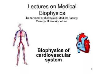
RA – right atrium RV – right ventricle LA – left atrium LV – left ventricle AV – atrioventricular valve Left AV – left atrioventricular valve Right AV - right atrioventricular valve SL – semilunar valve Common abbreviations used to refer to chambers: NB: No valves are present between major veins and atria. Hyperpressure leads to signs of congestion.
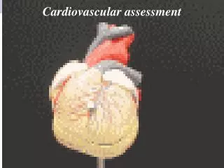
Topographical Landmarks of the Heart • Precordium – thepart of the ventral surface of the body overlying the heart and stomach and comprising the epigastrium and the lower median part of the thorax
Topographical Landmarks • Each area corresponds to one of the hearts 4 valves. • Aortic area - 2nd ICS to right of sternum (closure of the aortic valve loudest here). • Pulmonic area - 2nd ICS to left of sternum (closure of the pulmonic valve loudest here). • Tricuspid - 5th ICS left of sternal border (closure of tricuspid valve). • Mitral - 5th ICS left of the sternum just medial to MCL (closure of mitral valve). When cardiac output is increased as in anemia, anxiety, HTN, fever, the impulse may have greater force - inspect for lift or heave.
The first heart sound - systolic S1: Signals the closure of AV valves and the beginning of systole. Consists of mitral M1 and tricuspid T1 components. Is loudest at the apex Normal Heart Sounds
The second heart sound - diastolic S2: • Signals the closure of semilunar valves and the end of systole. • Consists of aortic A2 and pulmonic P2 components. • Is loudest at the base. • S1 & S2 correspond respectively to the familiar "lub dub" often used to describe the sounds.
Effect of respiration: MoRe to the Right heart Less to the Left • A split S2 – when the aortic valve closes significantly earlier than the pulmonic valve, you can hear the two components separately.
Other Heart Sounds • Extra Heart Sounds: • S3 • is the result of vibrations produced during ventricular filling. • is normally heard only in some children and young adults, but it is considered abnormal in older individuals. • S4 • is caused by the recoil of vibrations between the atria and ventricles following atrial contraction, at the end of diastole. • is rarely heard as a normal heart sound; usually it is considered indicative of further cardiac evaluation.
Other Heart Sounds • Murmurs: • are produced by vibrations within the heart chambers or in the major arteries from the back and forth flow of blood. • are classified as: • 1. Innocent, occurring in individuals with no anatomic or physiologic abnormality. • 2. Functional, occurring in individuals with no anatomic cardiac defect but with a physiologic abnormality such as anemia. • 3. Organic, occurring in individuals with a cardiac defect with or without a physiologic abnormality.
The conduction system of the heart consists of four structures: • 1. The sinoatrial(SA) node, located within the rig atrial wall near the opening of the superior vena cava • 2. The atrioventricular (AV) node, also located within the right atrium but near the lower end of the septum • 3. The atrioventricular bundle (bundle of His), which extends from the atrioventricular node along each side of the interventricular septum • 4. Purkinje fibers, which extend from the atrioventricular bundle into the walls of the ventricles. The electric impulses from this conduction system can be recorded on an electrocardiogram.
Electrocardiography (ECG) • records the electrical impulses generated from the heart muscle and provides a graphic illustration of the summation of these impulses and their sequence and magnitude.
The ECG waves • P wave represents the electric activity associated with the sinoatrial node and the spread of the impulse over the atria. It is a wave of depolarization. • QRS complex (wave) is composed of three separate waves: the Q wave, the R wave, and the S wave. They are all caused by currents generated when the ventricles depolarize before their contraction. Because ventricular depolarization requires septal and right and left ventricular depolarization, the electrical wave depicting these events is more complex than the smooth P wave. • P-R interval is measured from the beginning of the P wave to the beginning of the QRS complex. It is termed P-R instead of PQ because frequently the Q wave is absent. This interval represents the time that elapses from the begin Q-T intervalning of atrial depolarization to the beginning of ventricular depolarization.
The ECG waves • The T wave represents repolarization of the ventricles. The Q-T interval begins with the QRS complex and ends with the completion of the T wave. It represents ventricular j depolarization and repolarization. This interval varies with j the heart rate. The faster the rate, the shorter the Q-T interval. Therefore in children this interval is normally shorter than in adults. • The S-T segment is normally an isoelectric (flat) line that I connects the end of the S wave to the beginning of the T wave. • The T-P interval represents atrial and ventricular polarization in anticipation of the next cardiac cycle.
Pumping Ability • 4 to 6 L of blood per min throughout the body • Preload – venous return • Afterload – the opposing pressure the ventricles must generate to open aortic valve.
Developmental Considerations • Infants: • Transition from fetal circulation to postnatal circulation. By 9 months anatomical closure of foramen ovale occurs. • S1 and S2 sounds similarly on auscultation. Pulse rate 120/min. • Horizontal position of the heart (till 7-years-old).
Infants: Apex impulse is located at the 4th intercostal space 1 to 2 cm outward from left midclavicular line. Developmental Considerations
Developmental Considerations • The pregnant female: • By the end of pregnancy blood volume increases by 30 to 40 %. • Stroke volume and cardiac output are increased. • BP decreases due to vasodilation. • Pulse rate increases of 10 to 15 beats/min.
Developmental Considerations • An aging adult: • The incidence of CV diseases increases with age: coronary artery disease, HBP, heart failure.
Transcultural considerations • Smoking: widely spread in some societies. • HBP: Afro-Americans, Mexican-Americans and Native Americans have higher risk of hypertension. • Serum cholesterol: during childhood (4-19 yrs) Afro-American children have higher total cholesterol than Euro- and Mexican-Am. Children. This difference reverse during adulthood. • Obesity: more than 50% of Am. population are overweight. • Diabetes: the prevalence of diabetes increases in all groups in USA.
Physical Examination • Objectives: • Subjective data. • Health history data. • Preparation. • Inspection: general appearance, precordium. • Palpation: peripheral pulses, apical impulse. • Percussion. • Auscultation: heart sounds, murmurs. • Summary checklist.
Angina – an important cardiac symptom. “Clenched fist” sign is characteristic of angina. Chest pain: • Onset, location, character, aggravating and/or relieving factors • Character: crashing, stabbing, burning, vise-like. • Associated symptoms: sweating, ashen gray or pale skin, shortness of breath, nausea or vomiting, racing of heart, heart skips beat.
Subjective data Paroxysmal nocturnal dyspnea (PND) occurs with heart failure. Classically, the person awakens after 2 hrs. of sleep, arises, and flings open the window with the perception of needing fresh air. • Dyspnea: • Cause, onset, duration, affection by position, • Does shortness of breath interfere with activities of daily living? • Orthopnea: • Is the need to assume a more upright position to breathe. • Note the exact number of pillows used.
Hemoptysis is often a pulmonary problem, but also occurs with mitral stenosis Subjective data • Cough: duration, frequency, type, coughing up sputum (color, odor, blood tinged, aggravating and/or relieving factors. • Fatigue: onset, relation to time of day? • Cyanosis or pallor: occurs with myocardial infarction or low cardiac output.
Subjective data • Edema: • Swelling of legs or dependent body part due to increased interstitial fluid. • Onset, recent change, relation to time of day, relieving factors, associated symptoms. • Nocturia: • Occurs with heart failure in the person who is ambulatory during the day.
History taking. • Past cardiac history: • ! Last ECG, stress ECG, serum chilesterol measurements, other heart tests? • Family cardiac history: • Family history of hypertension, diabetes, heart problems, coronary artery disease (CAD), sudden death at younger age? • Personal habits (cardiac risk factors): nutrition, smoking, alcohol, exercise, drugs.
Additional history • For infants: mother’s health during pregnancy, feeding habits, growth, activity. • For children: growth, activity, any joint pains or unexplained fever, frequent headaches or nosebleedings, streptococcal infection (tonsillitis). • For pregnant female: any high PB during this or previous pregnancies, associated signs (weight gain, proteinuria), dizziness. • For aging adult: any symptoms of heart diseases (HTN, CAD) or COPD, any recent changes, medications (digitalis), side effects; environment.
Preparation • Bring to lab: • Watchwith second hand, • Stethoscope, • Marking pen and small centimeter ruler, • Alcohol swab (to clean endpiece). • Wear: • loose T-shirt or some other garment that allows for practice of physical assessment
Skin colour (cyanosis, pallor) and condition Any obvious bulging on anterior thorax at the left Edema Orhtopnea Inspection
Palpation • Palpate the apical impulse (the point of maximal impulse, or PMI): • Location: one intercostal space (usually 5th ICS) at left MCL, • Size: normally 1 cm 2 cm, • Amplitude: normally a shot, gentle tap, • Duration: short, normally occupies only first half of systole. • Ask the client “to exhale then hold it” or turn him to the left side.
Palpation • Palpate across the precordium for: • Other pulsations, • Thrill – palpable vibration due to strong heart murmur (like a purring cat), • Pericardial friction rubs are scratchy, high-pitched grating sounds, similar to pleural friction rubs, except that they are not affected by changes in respiration. • Accentuated S1 and S2. • A diffuse impulse (lift, heave).
Percussion • Is used to estimate approximately heart borders and configuration. • Recently is displaced by the chest x-ray or EchoCG. • Helps to detect heart enlargement Heart (cardiac) enlargement is due to increased ventricular volume or thickening of heart wall. Occurs with HTN, CAD, heart failure, cardiomyopathy
Auscultation • A Z-pattern is recommended. • Before beginning alert the person for long duration of procedure. • Begin with diaphragm endpiece and use the following routing: • Note the rate • the rhythm • Identify S1 and S2 • Listen for extra heart sounds • Listen for murmurs
Auscultation (cont.) Rhythm: • Regular • Irregular: • Synus arrythmia – common variation. Rate ↑ on inspiration and ↓ on expiration. • Regularly irregular • Irregularly irregular – no pattern to the sounds, beats come rapidly and at random intervals. • Pulse deficit – occurs with atrial fibrillation, heart failure, detects weak heart contractions.
Auscultation (cont.) • Identify S1 and S2 • Location and amplitude, • Correlation with peripheral pulses, PMI • Correlation with ECG waves • “Lub” or “dup” • Give description of origin. • Listen to sounds separately: accentuation, split (fixed, paradoxical).
Auscultation (cont.) • Extra heart sounds: • Midsystolic click • S3: normal, pathological (ventricular gallop) • S4: atrial gallop • Listen for murmurs: • Characteristics: timing, loudness, pitch, pattern, quality, location, radiation, posture
Grading murmurs • Grade I-VI: • Refers to the severity of a heart murmur (blowing, whooshing, or rasping sound), which is the result of vibrations caused by turbulent blood flow patterns. • Murmurs are classified ("graded") depending on their ability to be heard by the examiner. The grading is on a scale with grade I being barely detectable. • An example of a murmur description is a "grade II/VI murmur." (This means the murmur is grade 2 on a scale of 1 to 6).
Murmurs are classified according to their timing within the cardiac cycle. • Systolic Between S1and S2. • Diastolic Between S2 and S1). • Systolic ejection Begin after the first heart sound, attain a peak during midsystole, and terminate before the second heart sound. • Pansystolic or holosystolic During all of systole. • Pandiastolic or holodiastolic During all of diastole. • Prodiastolic Early diastolic. • Presystolic Late diastolic. • Continuous Continue through all of systole and all or part of diastole.
Conclusion • Function can be assessed to a large degree by findings in the history: shortness of breath (SOB), edema of ankles/legs, pain, pulse rate and rhythm; vital signs, signs and symptoms of oxygen deficit. • Location: Heart lies behind and to the left of the sternum. The upper portion or atria (BASE) lies to the back; the ventricles (APEX) points forward, the apex of the left ventricle actually touches the anterior chest wall near the left midclavicular line at or near the 5th left ICS. Known as point of maximal impulse (PMI) and is where apical beat is assessed. Impulse is a good index of heart size. • Landmarks for assessment: The precordium is the area on the anterior chest overlying the heart. Hearts sounds are heard throughout the precordium, but there are 4 major areas for examining heart sounds.