Download
1 / 35
350 likes | 499 Vues
This comprehensive overview of the cardiovascular system details the heart's structure, including its chambers, valves, and the conduction system. It explains how blood circulates through the pulmonary and systemic pathways, emphasizing the importance of mechanical and electrical properties. The learning outcomes prepare nurses to collect relevant subjective and objective data for patients with cardiovascular disorders, outline responsibilities for common diagnostic tests, and understand key concepts such as cardiac output, preload, and afterload.

E N D
Learning Outcomes • Describe the structure and function of the heart and vascular systems. • Discuss the mechanical and electrical properties of the heart.
Learning Outcomes • Identify subjective and objective assessment data to collect for patients with cardiovascular disorders. • Identify nursing responsibilities for common diagnostic tests and monitor for patients with cardiovascular disorders.
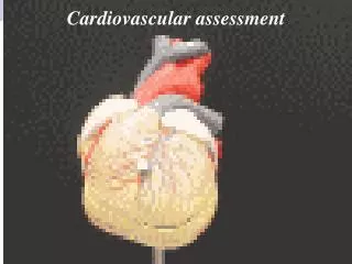
The Heart • Structure of the heart • Covered by pericardium • Parietal • Visceral (epicardium) • Epicardium • Outer heart layer • Myocardium • Middle heart layer
The Heart • Structure of the heart • Endocardium • Inner layer • Four hollow chambers • Two upper, atria • Two lower, ventricles • Divided by septum and atrioventricular (AV) valves
The Heart • Structure of the heart • Right atrium • Deoxygenated blood from superior and inferior vena cava • Into right ventricle, then pulmonary artery • Separated from right ventricle by tricuspid valve
The Heart • Structure of the heart • Left atrium • Oxygenated blood from lungs through pulmonary veins • Out of aorta to systemic circulation • Separated from left atria by bicuspid or mitral valve
Figure 15-3. Pulmonary and systemic circulation. The right side of the heart receives deoxygenated venous blood from the body and pumps it into the pulmonary circulation. The left side of the heart receives oxygenated blood from the pulmonary system and pumps it out to the peripheral circulation.
The Heart • Structure of the heart • Semilunar valves connect ventricles to great vessels. • Right—pulmonary valve • Left—aortic valve • S1 ("lub") sound • AV valves close as ventricles contract • S2 ("dub") sound • Semilunar valves close
The Heart • Structure of the heart • Coronary circulation • Blood to heart muscle • Left and right coronary arteries originate at base of aorta, encircle myocardium. • Cardiac veins • Drain blood into coronary sinus • Empty into right atrium of heart
The Heart • Conduction system • Electrical impulse, contraction without stimulation by nervous system • Sinoatrial (SA) node • Impulse • Travels through internodal pathways to AV node • Passes through bundle of His
The Heart • Conduction system • Impulse • Continues through right and left bundle branches • Out to Purkinje fibers in ventricular muscle walls • Action potential caused by movement of ions across cell membranes • Polarized (negative) • Depolarized (positive)
The Heart • Conduction system • All-or-nothing response • Repolarization immediately after depolarizaition • Refractory period • Cell resists stimulation • Protects heart from spasm, tetany
The Heart • Cardiac cycle • Diastole • Ventricular filling • Systole • Ventricles eject blood
The Heart • Cardiac output • Stroke volume (SV) • Volume of blood ejected with each contraction • Cardiac output (CO) • Amount of blood pumped by ventricles in one minute • CO = HR × SV
The Heart • Cardiac output • Cardiac reserve • Heart rate • Preload • Starling's law of the heart • Afterload • Contractility • Natural ability of cardiac muscle fibers to shorten during systole
ECG Video https://www.youtube.com/watch?v=7N4viIanngg https://www.youtube.com/watch?v=HJjTD0a8R00
Figure 15-6. The cardiac cycle. (A) Ventricular filling occurs during diastole (relaxation); (B) blood is pumped out of the heart to thepulmonary and systemic circulation during ventricular systole (contraction).
The Peripheral Vascular System • Network of blood vessels that carry blood to peripheral tissues and return it to the heart • Arteries • Veins • Capillaries
Arteries and Veins • Arteries • Carry blood away from heart • Divide into arterioles • Feed into beds of capillaries within organs, tissues • Veins • Carry blood toward heart • Venules feed into veins.
Arteries and Veins • Blood vessel structure • Layers • Tunica intima • Tunica media • Tunica externa • Veins have thinner walls. • Capillaries contain only one layer of tunica intima.
Arterial Circulation • Blood flow • Amount of blood transported • Peripheral vascular resistance (PVR) • Force of opposing blood flow • Determined by: • Blood viscosity • Vessel length • Vessel diameter
Arterial Circulation • Blood pressure (BP) • Force exerted by blood against wall of arteries • Systolic • Force exerted as heart contracts • Diastolic • Force exerted when heart is filling • Optimally less than 120/80 mm Hg in adults
Arterial Circulation • Factors affecting CO and PVR • Sympathetic nervous system • Parasympathetic stimulation • Kidneys • Temperatures • Chemicals, hormones, and drugs
Assessment • Health history • Chest pain • Shortness of breath (SOB) • Leg pain • Pillows to sleep • Medications • Lifestyle • Diet, alcohol use, exercise, smoking, drugs
Assessment • Physical examination • General appearance • Skin • Wounds • Pulses • Jugular vein distention • Edema • Breathing
The Older Adult • Subcutaneous tissue lost • Skin turgor more accurately assessed over sternum • Many experience early heartbeats. • Rate remains basically regular. • Increased incidence of chronic disease, medication use
Diagnostic Tests • Laboratory tests • Lipid profile • C-reactive protein • Serum cardiac markers • Serum cardiac hormones • Electrocardiography • Record of heart's electrical activity • Continual or intermittent based on patient needs
Diagnostic Tests • Imaging techniques • Sonography • Echocardiography • Transesophageal echocardiogram (TEE) • Stress echocardiogram • Vascular ultrasound • Radiography • Chest, abdominal x-rays
Diagnostic Tests • Imaging techniques • Angiography • Cardiac catheterization • CT scans • MRI
Diagnostic Tests • Imaging techniques • Nuclear scans • Multigated acquisition (MUGA) scan • Exercise perfusion imaging or stress tests