Download
1 / 67
710 likes | 1.04k Vues
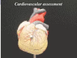
Assessment of the Cardiovascular System. Surface anatomy of the heart. The human heart is a cone-shaped, hollow, muscular organ located in the mediastinum between the lungs. It is approximately the size of an adult fist.

E N D
The human heart is a cone-shaped, hollow, muscular organ located in the mediastinum between the lungs. • It is approximately the size of an adult fist. • The heart rests on the diaphragm, tilting forward and to the left in the client's chest. • Each beat of the heart pumps approximately 60 mL of blood, or approximately 5 L/min. • During strenuous physical activity, the heart can double the amount of blood pumped to meet the increased oxygen needs of the peripheral tissues
RIGHT SIDE • The right atrium is a thin-walled structure that receives deoxygenated venous blood (venous return) from all peripheral tissues by way of the superior and inferior venae cavae and from the heart muscle by way of the coronary sinus. Most of this venous return flows passively from the right atrium, through the opened tricuspid valve, and to the right ventricle during ventricular diastole, or filling. The remaining venous return is actively propelled by the right atrium into the right ventricle during atrial systole, or contraction. • The right ventricle is a flat muscular pump located behind the sternum. The right ventricle generates enough pressure (approximately 25 mm Hg) to close the tricuspid valve, open the pulmonic valve, and propel blood into the pulmonary artery and the lungs. The workload of the right ventricle is light compared with that of the left ventricle because the pulmonary system is a low-pressure system, which imposes less resistance to flow
LEFT SIDE • After blood is reoxygenated in the lungs, it flows freely from the four pulmonary veins into the left atrium. Blood then flows through an opened mitral valve into the left ventricle during ventricular diastole. • When the left ventricle is almost full, the left atrium contracts, pumping the remaining blood volume into the left ventricle. With systolic contraction, the left ventricle generates enough pressure (approximately 120 mm Hg) to close the mitral valve and open the aortic valve. Blood is propelled into the aorta and into thesystemic arterial circulation
Blood is propelled from the aorta throughout the systemic circulation to the various tissues of the body; blood returns to the right atrium because of pressure differences. • The pressure of blood in the aorta of a young adult averages approximately 100 to 120 mm Hg, whereas the pressure of blood in the right atrium averages about 0 to 5 mm Hg. These differences in pressure produce a pressure gradient, with blood flowing from an area of higher pressure to an area of lower pressure. • The heart and vascular structures are responsible for maintaining these pressures
ATRIOVENTRICULAR VALVES • The AV valves separate the atria from the ventricles. • The tricuspid valve is composed of three leaflets and separates the right atrium from the right ventricle. • The mitral (bicuspid) valve is composed of two leaflets and separates the left atrium from the left ventricle • During ventricular diastole, the valves act as funnels and facilitate the flow of blood from the atria to the ventricles. During systole, the valves close to prevent the backflow (regurgitation) of blood into the atria
SEMILUNAR VALVES • There are two semilunar valves: the pulmonic valve and the aortic valve. • The pulmonic valve separates the right ventricle from the pulmonary artery. • The aortic valve separates the left ventricle from the aorta. • Each semilunar valve consists of three cuplike cusps, or pockets, around the inside wall of the artery. These cusps prevent blood from flowing back into the ventricles during ventricular diastole. During ventricular systole, these valves are open to permit blood flow into the pulmonary artery and the aorta
LEFT CORONARY ARTERY • The LCA divides into two branches: the left anterior descending (LAD) and the circumflex coronary artery (LCX). The LAD branch descends toward the anterior wall and the apex of the left ventricle. It supplies blood to portions of the left ventricle, ventricular septum, chordae tendineae, papillary muscle, and right ventricle. • The LCX descends toward the lateral wall of the left ventricle and apex. It supplies blood to the left atrium, the lateral and posterior surfaces of the left ventricle, and sometimes portions of the interventricular septum. In 45% of people, the LCX supplies the sinoatrial (SA) node, and in 10% of people it supplies the AV node. • Peripheral branches (diagonal and obtuse marginal) arise from the LAD and LCX and form an abundant network of vessels throughout the entire myocardium
RIGHT CORONARY ARTERY • The RCA originates from the right sinus of Valsalva, encircles the heart, and descends toward the apex of the right ventricle. • The RCA supplies the right atrium, right ventricle, and inferior portion ofthe left ventricle. • In most people (more than 50%), the RCA supplies the SA node and the AV node. • Considerable variation in the branching pattern of the coronary arteries exists among individuals
The cardiac conduction system is composed of specialized tissue capable of rhythmic electrical impulse formation. It can conduct impulses much more rapidly than other cells located in the myocardium. • The SA node, located at the junction of the right atrium and the superior vena cava, is considered the main regulator of heart rate. The SA node is composed of pacemaker cells, which spontaneously initiate impulses at a rate of 60 to 100 times per minute and myocardial working cells, which transmit the impulses to the surrounding atrial muscle • An impulse from the SA node initiates the process of depolarization and hence the activation of all myocardial cells. The impulse travels through both atria to the atrioventricular (AV) node located in the junctional area. After the impulse reaches the AV node, conduction of the impulse is delayed briefly. This delay allows the atria to contract completely before the ventricles are stimulated to contract. The intrinsic rate of the AV node is 40 to 60 beats/min.
The Bundle of His is a continuation of the AV node and is located in the interventricular septum. It divides into the right and left bundle branches. • The bundle branches extend downward through the ventricular septum and fuse with the Purkinje fiber system. • The Purkinje fibers are the terminal branches of the conduction system and are responsible for carrying the wave of depolarization to both ventricular walls. Purkinje fibers can act as an intrinsic pacemaker, but their discharge rate is only 20 to 40 beats/min. • Thus these intrinsic pacemakers seldom initiate an electrical impulse
Mechanical properties of the heart • The electrical and mechanical properties of cardiac muscle determine the function of the cardiovascular system. The heart is able to adapt to various pathophysiologic conditions (e.g., stress, infections, and hemorrhage) to maintain adequate blood flow to the various body tissues. • Blood flow from the heart into the systemic arterial circulation is measured clinically as cardiac output (CO), the amount of blood pumped from the left ventricle each minute. CO depends on the relationship between heart rate (HR) and stroke volume (SV); it is the product of these two variables: • Cardiac output = Heart rate x Stroke volume
CARDIAC OUTPUT AND CARDIAC INDEX • Cardiac output (CO) is the volume of blood (in liters) ejected by the heart each minute. In adults, the CO ranges from 4 to 7 L/min. • Because cardiac output requirements vary according to body size, the cardiac index is calculated to adjust for differences in body size. • The cardiac index can be determined by dividing the CO by the body surface area. • The normal range is 2.7 to 3.2 L/min/m2 of body surface area
HEART RATE • Heart rate refers to the number of times the ventricles contract each minute. The normal resting heart rate for an adult is between 60 and 100 beats/min. Increases in heart rate increase myocardial oxygen demand. • Heart rate is extrinsically controlled by the autonomic nervous system, which adjusts rapidly when necessary to regulate cardiac output. The parasympathetic system slows the heart rate, whereas sympathetic stimulation has an excitatory effect. An increase in circulating endogenous catecholamine (e.g., epinephrine and norepinephrine) usually causes an increase in heart rate, and vice versa. • Other factors, such as the central nervous system (CNS) and baroreceptor (pressoreceptor) reflexes, influence the effects of the autonomic nervous system on heart rate. Pain, fear, and anxiety can increase heart rate. • The baroreceptor reflex acts as a negative-feedback system. If a client experiences hypotension, the baroreceptors in the aortic arch sense a lessened pressure in the blood vessels. A signal is relayed to the parasympathetic system to have less of an inhibitory effect on the sinoatrial (SA) node; this results in a reflex increase in heart rate
STROKE VOLUME • Stroke volume is the amount of blood ejected by the left ventricle during each systole. Severalvariables influence stroke volume and, ultimately, CO. • These variables include heart rate, preload, afterload, and contractility
PRELOAD • Preload refers to the degree of myocardial fiber stretch at the end of diastole and just before contraction. The stretch imposed on the muscle fibers results from the volume contained within the ventricle at the end of diastole. Preload is determined by left ventricular end-diastolic (LVED) volume. • An increase in ventricular volume increases muscle fiber length and tension, thereby enhancing contraction and improving stroke volume. • This statement is derived from Starling's law of the heart: the more the heart is filled during diastole (within limits), the more forcefully it contracts. • However, excessive filling of the ventricles results in excessive LVED volume and pressure and a decreased cardiac output
AFTERLOAD. • Afterload is the pressure or resistance that the ventricles must overcome to eject blood through the semilunar valves and into the peripheral blood vessels. The amount of resistance is directly related to arterial blood pressure and the diameter of the blood vessels. • Impedance, the peripheral component of afterload, is the pressure that the heart must overcome to open the aortic valve. The amount of impedance depends on aortic compliance and total systemic vascular resistance, a combination of blood viscosity and arteriolar constriction. • A decrease in stroke volume can result from an increase in afterload without the benefit of compensatory mechanisms
CONTRACTILITY • Contractility also affects stroke volume and CO. • Myocardial contractility is the force of cardiac contraction independent of preload. • Contractility is increased by factors such as sympathetic stimulation and calcium release. • Factors such as hypoxia and acidemia decrease contractility
Blood Pressure • Blood pressure is the force of blood exerted against the vessel walls. • The blood pressure in the arterial system is determined primarily by the quantity of blood flow or cardiac output (CO), as well as by the resistance in the arterioles: Blood pressure = Cardiac output x Peripheral vascular resistance
Blood Pressure Regulation • Autonomic nervous system • Baroreceptors • Chemoreceptors • Renal system • Endocrine system • External factors also affect BP
Venous System • Structure: a series of veins located adjacent to the arterial system • Function: completes the circulation of blood by returning blood from the capillaries to the right side of the heart • Cardiovascular changes in the older adult: only evident when the person is active or under stress
Assessment Techniques • History • Demographic data • Family history and genetic risk • Personal history • Diet history • Socioeconomic status
Modifiable Risk Factors • Cigarette smoking • Physical inactivity • Obesity • Psychological factors • Chronic disease
Pain or Discomfort • Pain or discomfort can result from ischemic heart disease, pericarditis, and aortic dissection. • Chest pain can also result from noncardiac conditions such as pleurisy, pulmonary embolus, hiatal hernia, and anxiety. (Continued)
Pain or Discomfort (Continued) • Terms such as discomfort, heaviness, pressure, indigestion, aching, choking, strangling, tingling, squeezing, constricting, or vise-like are all used to describe pain. • Women often do not experience pain in the chest but rather feelings of discomfort or indigestion.
Pain Assessment • Onset • Manner of onset • Duration • Frequency • Precipitating factors • Location • Radiation (Continued)
Pain Assessment(Continued) • Quality • Intensity, which can be graded from 0 to 10, associated symptoms, aggravating factors, and relieving factors
Dyspnea • Can occur as a result of both cardiac and pulmonary disease • Difficult or labored breathing experienced as uncomfortable breathing or shortness of breath • Dyspnea on exertion (DOE) • Orthopnea: dyspnea when lying flat • Paroxysmal nocturnal dyspnea after lying down for several hours
Other Manifestations • Fatigue • Palpitations • Weight gain • Syncope • Extremity pain
Physical Assessment • General appearance • The nurse assesses the following areas: general build and appearance, skin color, distress level, level of consciousness, shortness of breath, position, and verbal responses. • Clients with chronic heart failure may appear malnoutrished, thin, and cachectic. • Latent signs of severe heart failure are ascites, jaundice, and anasarca (generalized edema) as a result of prolonged congestion of the liver. • Heart failure may cause fluid retention, and clients may have engorged neck veins and generalized dependent edema. • Coronary artery disease is suspected in clients with yellow, lipid-filled plaques on the upper eyelids (xanthelasma) or ear-lobe creases. • Clients with poor cardiac output and decreased cerebral perfusion may experience mental confusion, memory loss, and slowed verbal responses
Physical Assessment • Integumentary system • Skin color • If there is normal blood flow or adequate perfusion to a given area in light-colored skin, it appears pink, perhaps rosy incolor, and it is warm to the touch. Decreased flow is depicted as cool, pale, and moist skin. Pallor is characteristic of anemia and can be seen in areas such as the nail beds, palms, and conjunctival mucous membranes • A bluish or darkened discoloration of the skin and mucous membranes in Caucasians is referred to as cyanosis. This condition results from an increased amount of deoxygenated hemoglobin. Dark-skinned individuals may express cyanosis as a graying of the same tissues
Physical Assessment • Central cyanosis involves decreased oxygenation of the arterial blood in the lungs and appears as a bluish tinge of the conjunctivae and the mucous membranes of the mouth and tongue. Central cyanosis may indicate impaired lung function or a right-to-left shunt found in congenital heart conditions. Because of impaired circulation, there is a marked desaturation of hemoglobin in the peripheral tissues, which produces a bluish or darkened discoloration of the nail beds, earlobes, lips, and toes. • Peripheral cyanosis occurs when blood flow to the peripheral vessels is decreased by peripheral vasoconstriction. The clamping down of the peripheral blood vessels results from a low cardiac output or an increased extraction of oxygen from the peripheral tissues. Peripheral cyanosis localized in an extremity is usually a result of arterial or venous obstruction
Physical Assessment • Skin temperature • Skin temperature can be assessed for symmetry by touching different areas of the client's body (e.g., arms, hands, legs, and feet) with the dorsal surface of the hand or fingers. • Decreased blood flow results in decreased skin temperature. Skin temperature is lowered in several clinical conditions, including heart failure, peripheral vascular disease, and shock
Physical Assessment • Extremities • The nurse assesses the client's hands, arms, feet, and legs for skin changes, vascular changes, clubbing, capillary filling,and edema. • Skin mobility and turgor are affected by the fluid status of the client. • Dehydration and aging reduce skin turgor, and edema decreases skin mobility. • Vascular changes in an affected extremity may include paresthesia, muscle fatigue and discomfort, numbness, pain, coolness, and loss of hair distribution from a reduced blood supply
Physical Assessment • Blood pressure • Normal blood pressure in adults older than 45 years of age ranges from 90 to 140 mm Hg for systolic pressure and from 60 to 90 mm Hg for diastolic pressure. • A blood pressure that exceeds 135/85 mm Hg increases the workload of the left ventricle and oxygen consumption. • A blood pressure less than 90/60 mm Hg may be inadequate for providing proper and sufficient nutrition to bodycells. In certain circumstances, such as shock and hypotension, the Korotkoff sounds are less audible or are absent. In these cases the nurse might palpate the blood pressure, use an ultrasonic device (Doppler device), or obtain a direct measurement by arterial catheter
Physical Assessment • Venous and arterial pulses: central and jugular venous pressures, and jugular venous distention • The nurse observes the venous pulsations in the neck to assess the adequacy of blood volume and central venous pressure (CVP). The nurse can assess jugular venous pressure (JVP) to estimate the filling volume and pressure on the right side of the heart. • The right internal jugular vein is usually used to estimate JVP
Precordium • Assessment of the precordium (area over the heart) involves: • Inspection • Palpation • Percussion • Auscultation • Normal heart sounds • Paradoxical splitting • Gallops and murmurs • Pericardial friction rub
Normal heart sounds • The first heart sound (S1) is created by the closure of the mitral and tricuspid valves (atrioventricular valves). • When auscultated, the first heart sound is softer and longer; it is of a low pitch and is best heard at the lower left sternal border or the apex of the heart. • It may be identified by palpating the carotid pulse while listening. S1 marks the beginning of ventricular systole and occurs right after the QRS complex on the electrocardiogram (ECG). • The first heart sound can be accentuated or intensified in conditions such as exercise, hyperthyroidism, and mitral stenosis. • A decrease in sound intensity occurs in clients with mitral regurgitation and heart failure.
Normal heart sounds • The second heart sound (S2) is caused mainly by the closing of the aortic and pulmonic valves (semilunar valves). • S2 is characteristically shorter. It is higher pitched and is heard best at the base of the heart at the end of ventricular systole. • The splitting of heart sounds is often difficult to differentiate from diastolic filling sounds (gallops). A splitting of S1 (closure of the mitral valve followed by closure of the tricuspid valve) occurs physiologically because left ventricular contraction occurs slightly before right ventricular contraction. • However, closure of the mitral valve is louder than closure of the tricuspid valve, so splitting is often not heard. • Normal splitting of S2 occurs because of the longer systolic phase of the right ventricle. Splitting of S1 and S2 can be accentuated by inspiration (increased venous return), and it narrows during expiration.
Abnormal heart sounds PARADOXICAL SPLITTING. • Abnormal splitting of S2 is referred to as paradoxical splitting and is characteristic of a wider split heard on expiration. • Paradoxical splitting of S2 is heard in clients with severe myocardial depression that causes early closure of the pulmonic valve or a delay in aortic valve closure. • Such conditions include myocardial infarction, left bundle branch block, aortic stenosis, aortic regurgitation, and right ventricular pacing.
Abnormal heart sounds GALLOPS AND MURMURS. • GALLOPS. Diastolic filling sounds (S3) and (S4) are produced when blood enters a noncompliant chamber during rapid ventricular filling. • The third heart sound (S3) is produced during the rapid passive filling phase of ventricular diastole when blood flows from the atrium to a noncompliant ventricle. The sound arises from vibrations of the valves and supporting structures. • The fourth heart sound (S4) occurs as blood enters the ventricles during the active filling phase at the end of ventricular diastole. • S3 is termed ventricular gallop, and S4 is referred to as atrial gallop. • These sounds can be caused by decreased compliance of either or both ventricles. • The nurse can best hear left ventricular diastolic filling sounds with the client on his or her left side. The bell of the stethoscope is placed at the apex and at the left lower sternal border during expiration.