Comprehensive Cardiovascular Assessment Guide
470 likes | 600 Vues
Learn how to assess specific heart problems including chest pain, tachycardia, fatigue, dyspnea, and more. Gather subjective and objective data accurately for a thorough examination.

Comprehensive Cardiovascular Assessment Guide
E N D
Presentation Transcript
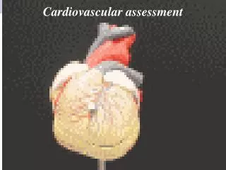
Cardiovascular Assessment Dr. Zyad Saleh
Subjective Data • specific heart problems: • Chest pain • cardiac, pulmonary, muscular, or gastrointestinal • Sensation of squeezing around the heart; a steady, severe pain; and a sense of pressure. • radiate to the left shoulder and down the left arm or to the jaw. • Diaphoresis and pain worsened by activity • dyspnea, diaphoresis, pallor, nausea, palpitations, or tachycardia.
Tachycardia and Palpitations (beat faster) • Tachycardia increase cardiac output. • Palpitations abnormality of the heart’s conduction (arrhythmia) or increase cardiac output by Tachycardia
Fatigue • Fatigue may result from compromised cardiac output. • Fatigue related to decreased cardiac output is worse in the evening or as the day progresses,
Difficulty of breathing (dyspnea) or shortness of breathing • Dyspnea cardiac or pulmonary disorders • Dyspnea may occur at rest, during sleep, or with exertion. • Orthopnea is the need to sit more upright to breath easily • paroxysmal nocturnal dyspnea (wake up from dyspnea) heart failure due to redistribution of fluid from the ankles to the lungs when one lies down at night.
Cough up with mucous • Fluid accumulation in the lungs from heart failure white to pink-tinged sputum. • Dizziness • Dizziness decreased blood flow to the brain
Wake up with an urgent need to urinate (nocturia) • Increased renal perfusion • Swelling edema in feet, ankles, and legs • Edema in both lower extremities at night • dependent areas of the body.
Heartburn • Gastrointestinal pain may occur after meals and is relieved with antacids. • Cardiac pain may occur anytime, is not relieved with antacids, and worsens with activity.
INSPECTION: Neck Vessels Observe the jugular venous pulse
INSPECTION: Neck Vessels • Observe the jugular venous pulse • The jugular venous pulse is not normally visible with the client sitting upright. (position fully distends the vein) • pulsations may or may not be discernible. • increased central venous pressure
Evaluate jugular venous pressure • The jugular vein should not be distended, bulging, or protruding at 45 degrees or greater • Right side heart failure
Auscultation and Palpation • Auscultate the carotid arteries if you suspect cardiovascular disease. • No blowing or swishing or other sounds are heard. • Pulses are equally strong; a 2+, with no variation in strength • Contour is smooth and rapid on upstroke and slower on downstroke. • Pulse Amplitude Scale 0 = Absent 1+ = weak 2+ = Normal 3+ = Increased 4+ = Bounding
occlusive arterial disease A bruit, a blowing or swishing sound by turbulent blood flow through a narrowed vessel Pulse inequality arterial constriction or occlusion in one carotid. Weak pulses hypovolemia, shock, or decreased cardiac output. A bounding, firm pulse may indicate hypervolemia or increased cardiac output. Variations in strength from beat to beat or with respiration abnormal A delayed upstroke aortic stenosis.
Palpate the carotid arteries. • Arteries are elastic and no thrills • Loss of elasticity atherosclerosis • Thrills a narrowing of the artery.
INSPECTION: Heart (Precordium) is the portion of the body over the heart and lower chest Inspect pulsations.
Aortic area at the right second intercostal space–S2 is louder than S1 • Pulmonic area :at the left second intercostal space–S2 is louder than S1 • Erb’s point: at the left third intercostal space–S1 and S2 are heard equally • Tricuspid area at the left fourth intercostal space–S1 is louder than S2 • Apex:the left fifth intercostal space at the midclavicular line–S1 is louder than S2
INSPECTION: Heart (Precordium) • Inspect pulsations. • The apical impulse may or may not be visible. • it would be in the mitral area (left mid-clavicular line, fifth or fourth intercostal space) • Apical impulses result of the left ventricle moving outward during systole.
PALPATION • Palpate the apical impulse. • The apical impulse is palpated in the mitral area • Size 1-2 cm • Amplitude small like a gentle tap. • The duration is brief, lasting through the first 2/3 of systole or less • obese clients may not be palpable.
The apical impulse may be impossible to palpate in clients with pulmonary emphysema. Cardiac enlargement large size displaced, more forceful, or of longer duration.
Palpate for abnormal pulsations. • No pulsations or vibrations are palpated in the areas of the apex, left sternal border, or base. • A thrill or a pulsation is usually associated with murmur.
AUSCULTATION • Auscultate heart rate and rhythm. • Rate = regular 60 to 100 • Bradycardia • Tachycardia • Arrhythmia • Cause Decrease cardiac output or emboli
If you detect an irregular rhythm, auscultate for a pulse rate deficit. • The radial and apical pulse rates should be identical. • pulse rate deficit (difference between apical pulse and peripheral pulse arrhythmia
Auscultate to identify S1 and S2. • The first heart sound (S1) is the result of closure of the AV valves: the mitral and tricuspid valves. • S1 (“lub”) is usually heard as one sound but may be heard as two sounds • two sounds, the first component represents mitral valve closure (M1); the second component represents tricuspid closure (T1). • heard best at the apex (left MCL, fifth ICS).
Auscultate to identify S1 and S2. • second heart sound (S2) results from closure of the semilunar valves (aortic and pulmonic) • two sounds, the first component represents aortic valve closure (A2) and the second component represents pulmonic valve closure (P2). • heard best at the base of the heart.
Auscultate to identify S1 and S2. • First heart sound = S1 or lub • Second heart sound = S2 or dubb • two sounds make up the cardiac cycle of systole and diastole. • S1 starts systole, and S2 starts diastole. The space between S1 and S2 (systolic pause) is of short duration • The space between S2 and the start of another S1 (diastolic pause) is of longer duration.
S1 corresponds with each carotid pulsation and is loudest at the apex of the heart. S2 immediately follows after S1 and is loudest at the base of the heart. diminished, varying, or split
An accentuated S1 sound is louder than an S2. This occurs when the mitral valve is wide open and closes quickly. Diminished S1: S1 sound is softer than the S2 sound. This occurs when the mitral valve is not fully open at the time of ventricular contraction and valve closing.
a split S1 occurs when the left and right ventricles contract at different times (asynchronous ventricular contraction). Varying S1 occurs when the mitral valve is in different positions when contraction occurs.
An accentuated S2 means that S2 is louder than S1. conditions in which the aortic or pulmonic valve has a higher closing pressure. A diminished S2 means that S2 is softer than S1. conditions in which the aortic or pulmonic valves have decreased mobility.
Auscultate for extra heart sounds. • Auscultate during the systolic and diastolic pause • S3 heart sound heard at the beginning of the diastolic pause • S4 heart sound heard at the end of the diastole
Extra Heart Sound S3 and S4 are referred to as diastolic filling sounds, or extra heart sounds, which result from ventricular vibration secondary to rapid ventricular filling. Description: Both sounds are low frequency and thus best heard with the bell of the stethoscope. Location: Usually best heard over apex with patient in the left lateral position
THIRD HEART SOUND S3 Results from increased atrial pressure leading to increased flow rates, as seen in congestive heart failure, which is the most common cause of a S3. May be normal physiological finding in patients less than age 40,pregnancy,athlatist or fever.
FOURTH HEART SOUND S4 Seen in patients with stiffened left ventricles, resulting from conditions such as hypertension, aortic stenosis, ischemic or hypertrophic cardiomyopathy.
Auscultate for murmurs. • A murmur a swishing sound caused by turbulent blood flow through the heart valves or great vessels. • increased blood velocity, structural valve defects, valve malfunction, and abnormal chamber openings • Thrills are superficial vibratory sensations felt on the skin overlying an area of turbulence
Systolic Murmurs These are ejection murmurs May be caused by turbulence across the aortic or pulmonic valves if they are stenosed May be caused by turbulence across the mitral or tricuspid valves if they are incompetent (regurgitant) The murmur falls between S1 and S2 Sounds like, LUB-shhh-dub
Diastolic Murmurs Mitral and tricuspid stenosis can cause a diastolic murmur. Aortic or pulmonic regurgitation can cause a diastolic murmur. Sounds like this: Lub-dub-shhh
auscultate with the client in different positions because some murmurs occur or subside according to the client’s position.