Hierarchical Image Segmentation for Identifying Stroke Regions
130 likes | 282 Vues
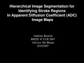
Hierarchical Image Segmentation for Identifying Stroke Regions In Apparent Diffusion Coefficient (ADC) Image Maps. Anthony Bianchi BRITE @ UCR 2007 Advisor: Bir Bhanu 8/24/2007. Outline. Background How it Happens The Images Why Automatic is Needed Process Flowchart

Hierarchical Image Segmentation for Identifying Stroke Regions
E N D
Presentation Transcript
Hierarchical Image Segmentation for Identifying Stroke Regions In Apparent Diffusion Coefficient (ADC) Image Maps Anthony Bianchi BRITE @ UCR 2007 Advisor: Bir Bhanu 8/24/2007
Outline • Background • How it Happens • The Images • Why Automatic is Needed • Process • Flowchart • Automatic Thresholding • Connected Components • Example • Results • Two Patients • Findings • Conclusion • Acknowledgments
Background • Stroke: 1/4000 live births • Arterial-ischemic stroke, 73% Arterial Ischemic Stroke Cerebral arterial thrombosis: possible postnatal etiology of AIS. AIS Focal Lesion
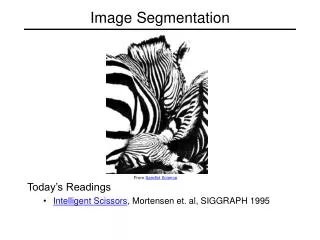
Apparent Diffusion Coefficient (ADC) Image Maps ≤ 5 days • An ADC image map measures the diffusion of water. If the diffusion is low the grayscale value is low. 5 days ≥ 5 days 25 days
Why is Automatic Segmentation Needed? • Currently manual segmentations is time intensive and inaccurate. Manual segmentations can very over 30% from one person to the next, and can take hours per patient. • An automatic segmentation algorithm will be repeatable, and will take minutes per patient. • We are currently working with LLUMC. They would like to use this segmentation to classify stroke victims into mild, moderate, and severe. They will use these labels to accept patients for stem cell trials.
The Process Find Image to be Segmented Find Threshold Automatically Split Image Using Threshold Higher Region Lower Region Find Connected Components That Satisfy Second threshold Find Connected Components That Satisfy Second threshold # Regions > 1 # Regions > 1 YES YES NO NO Close and Fill Images Separation Results
Automatic Thresholding: Otsu’s method 2w (t) = q1(t) 21(t) + q2(t) 22(t) Sum of Probability in Group 1 Sum of Probability in Group 2 Variance Group 2 Within Group Variance Variance Group 1 2w (t) = q1(t) 21(t) + q2(t) 22(t) • We test every threshold to find the smallest Within Group Variance. • A recursive form of the above equation is implemented to cut down computation time. Threshold Found 176
Connected Components Mask • A mask gets sent through the image. Each pixels is evaluated by the mask to see if it has a neighboring pixel. If there is a neighboring pixel the selected pixel gains the same label of that pixel. If no neighboring pixel is found a new label is created for that pixel. Example of Connected Components
Example of the Process Find object to be segmented. Threshold found 149 Object > 50% found Threshold found 98 Object found closed and filled
Patient 1 RED = Damage GREEN = Area RED = Damage GREEN = Area Patient 2
Results • For patient 1 the automatically segmented data gave a total damage of 14.7%, while manually segmented images gave a total of 17.7% damage. • The reason for the difference between the manual and automatic segmented is because the area used in finding total area in the automatic segmented included spinal fluid. This fluid can be found by the automatic method and can be removed. • For patient 2 we found the damaged area to be 3.5%, and the manual segmentation gave a 3.6% result. For patient two the manual segmentation included the cerebral spinal fluid, which was included in the area.
Conclusion • The experiments show that the automatic method has small differences compared to the manually segmented images. But, it is effective and consistent in finding the damage area in the ADC images. • Hypoxic-Ischemic Encephalopathy is another type of stroke that happens every 1/1000 live births. These injuries are diffused through the brain unlike the AIS patients. This segmentation method should be able to detect this type of stroke. • The next step is trying to use this method on different MRI types such as T2 image maps. • A 3D approach could give better results, because it could connect the structure from slide to slide.
Acknowledgments • I would like to thank: • Dr. Bhanu for his guidance. • Jacqueline Coats, Andy Obenaus, and Stephen Ashwal (from LLUMC) for data and useful information. • BRITE advisors for the opportunity. • Friends & Family for Support.