Download
1 / 75
1.01k likes | 2.86k Vues
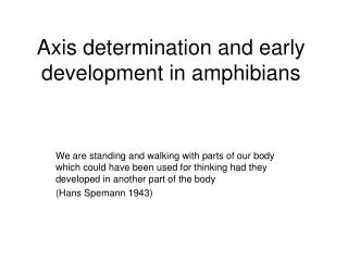
Chapter 07-Amphibians & Fish Early development and axis formation. 7.1 Formation of the parallel arrays of microtubules extend along the vegetal hemisphere along the future dorsal-ventral axis. These microtubules allow the cortical cytoplasm to rotate with respect to the inner cytoplasm.

E N D
Chapter 07-Amphibians & Fish Early development and axis formation
7.1 Formation of the parallel arrays of microtubules extend along the vegetal hemisphere along the future dorsal-ventral axis These microtubules allow the cortical cytoplasm to rotate with respect to the inner cytoplasm
7.1C Reorganization of cytoplasm in the newly fertilized frog egg (Part 1) animal Gastruation will begin in the gray crescent, region opposite of sperm entry (Ventral ) Dorsal First cleavage (80%) Vegetal
7.1D Reorganization of cytoplasm in the newly fertilized frog egg (Part 2)
Figure 10.3 Cleavage of a Frog Egg Cleavage in amphibians The first division begins at the animal pole and slowly extends down cleavage Figure 10.3A 4 micromeres: quick division • Two major functions: • Permit cell migration • Prevents the contact of the vegetal cells destines to become endoderm with those cells fated to give rise to the skin and nerves 4 macromeres: quick division The vegetal hemisphere ultimately contains larger and fewer blastomeres than the animal half. or “size discrepancy” Figure 7.2,3 Scanning Electron Micrographs of the Cleavage of a Frog Egg
Figure 7.4 Depletion of EP-cadherin mRNA in the Xenopus Oocyte While the cells are dividing in the oocyte, numberous cell adhesion molecules keep the blastomeres together. EP-cadherin: a maternal supply Antisense oligonucleotides Control The adhesion between the blastomeres is dramatically reduced, resulting in the obliteration of the blastocoel
Figure 10.25 Model of the Mechanism by which the Disheveled Protein Stabilizesb-catenin in the Dorsal Portion of the Amphibian Egg Translocated dorsally released (GSK-3b+Axin1(2)+APC) complex for b-catenin degradation Dsh protein is the inhibitor of GSK-3b
10.24 Model of the mechanism by which the Disheveled protein stabilizes -catenin in the dorsal portion of the amphibian egg (Part 1)
10.24 Model of the mechanism by which the Disheveled protein stabilizes -catenin in the dorsal portion of the amphibian egg (Part 2)
b-Catenin Tcf/Lef Tcf/Lef Tcf/Lef Transcription (c-Jun,fra-1,c-myc & cyclin D1 etc.) The Wnt signaling pathway in normal and cancer development Wnt Fz(frizzled) DSH GBP Akt GSk-3b GSk-3b GSk-3b APC APC APCmt Axin b-Catenin Axin Axinmt P P b-Catenin b-Catenin b-Catenin P Oncogenic b-Catenin P Stablization degradation b-Catenin b-Catenin n n b-Catenin CBP ICAT Groucho/TLE Chibby Repression Transcription (c-Jun,fra-1,c-myc & cyclin D1 etc.) Embryonic cell Differentiated cell Tumor cell
Cytoplasmic determinants in the egg.The unfertilized egg cell has molecules in its cytoplasm, encoded by the mother’s genes, that influence development. Many of these cytoplasmic determinants, like the two shown here, are unevenly distributed in the egg. After fertilization and mitotic division, the cell nuclei of the embryo are exposed to different sets of cytoplasmic determinants and, as a result, express different genes. a Maternal supply: Cytoplasmic Determinants and Cell-Cell Signals in Cell Differentiation Cytoplasmic determinants in the cytoplasm of the unfertilized egg Regulate the expression of genes in the zygote that affect the developmental fate of embryonic cells Unfertilized egg cell Sperm Molecules of another cyto- plasmic deter- minant Molecules of a a cytoplasmic determinant Fertilization Nucleus Sperm Zygote (fertilized egg) Mitotic cell division Two-celled embryo
Figure 7.5 Fate Maps of the Blastula of the Frog Xenopus laevis Amphibians Gastrulation The three germ layers Fate mapping has shown that cells of the Xenopus blastula have different fates depending on whether they are located in the deep or The superfical layers. The mesodermal precursors exist mostly in the deep layer of cells (the internal cytoplasm around the equator), while the ectoderm (the animal hemisphere surface) and endoderm (the vegetal hemisphere surface) arise from the superficial layer on the surface. Animal equator Vegetal
early Mid- endoderm Figure 7.6 Cell Movements During Frog Gastrulation (primitive gut) Sperm entry Fig. 10.8 Dorsal Marginal Zone cells Figure 7.7 Surface View of An Early Dorsal Blastopore Lip of Xenopus
10.9 A graft of cells from the dorsal marginal zone of a salamander embryo sinks into a layer of endodermal cells and forms a blastopore-like groove
7.7 Surface view of an early dorsal blastopore lip of Xenopus dorsal blastopore lip
10.11 Epiboly of the ectoderm (Part 2) Invaginate
Figure 7.11 Protocadherin expression separates axial and paraxial mesoderm
Figure 7.14 Spemann’s Demonstration of Nuclear Equivalence in Newt Cleavage Organizer for axis formation Spemann’s experiment I His baby girl’s hair Spemann’s experiment: early amphibian nuclei were geneticall identical and that each cell was able to give rise to an entire animal. Early amphibian nuclei were genetically identical and that each cell was capable of giving rise to an entire organism
Figure 7.15 Asymmetry in the Amphibian Egg Spemann’s experiment II: Only the blastomere containing the gray crescent develops normally The gary crescent region gives rise to the cells that initiate gastrulation. These cells form the dorsal lip of the blastopore. Fig. 10-2 The movements of fertilized amphibian eggs expose a gray crescent shaped are of cytoplasm in the region directly opposite the sperm entry point. No dorsal structures
(interspcies transplantation) Figure 10.18 Determination of Ectoderm During Newt Gastrulation Spemann’s experiment III: He found the cells of the early gastrula were uncommitted (regulative/conditional development), but the fates of late gastrula cells were determined(autonomous/independent/mosaic development). 可朔性高 uncommitted X 可朔性低 autonomous Why ? √ Organizer
Organization of a secondary axis by dorsal blastopore lip tissue (Part 1)
Organization of a Secondary Axis by Dorsal Blastopore Lip Tissue Spemann’s ( and Mangold) experiment IV:interspcies transplantation-Triturus taeniatus (pigmented; donor) vs Triturus cristatus (nonpigmented; host) . They found that all the tissue in the early gastrula, only one has its fate determined. It is the dorsal blastopore lip tissue (derived from the gray crescent cytoplasm)-the Organizer
10.19 Organization of a secondary axis by dorsal blastopore lip tissue (Part 2)
Figure 7.16 Determination of ectoderm during newt gastrulation
Figure 7.17 Organization of a secondary axis by dorsal blastopore lip tissue
Figure 10.10 Twin Blastopores Produced by Rotating Dejellied Xenopus Eggs Ventral Side Up at the Time of First Cleavage Although the sperm entry is not needed to induce the movements in the egg cytoplasm. The centrifuged eggs and two dorsal blastopore lips formed, leading to the formation of twin larvae Also see in zebrafish Siamese
Figure 7.18 Summary of Experiments by Nieuwkoop and by Nakamura and Takasaki, Showing Mesodermal Induction by Vegetal Endoderm Mechanisms of axis determination-Nieuwkoop center; b-catenin The experiments of Pieter Nieukoop and Osamu Nakamura Removed b-catenin The signal (TGF-b & FGFs) induces the marginal cells to become mesoderm
Figure 10.21 Transplantation Experiments on 64-cell Amphibian Embryos Nieuwkoop center demonstration The transplantation experiments of Gimlich & Gerhart (1984) blastopore √ √ √ The three vegetal blastomeres opposite the point of sperm entry are able to indurce the formation of the dorsal lip of the blastopore and a complete dorsal axis. also see Fig. 10-23
Figure 7.19 Transplantation and recombination experiments on Xenopus embryos
Figure 10.23 The Regional Specificity of Mesoderm Iinduction Can Be Demonstrated by Recombining Blastomeres of 32-Cell Xenopus Embryos (also see Fig. 10-11, 22C) Sperm entrance The dorsalmost vegetal cells induced the animal pole cells to become dorsal mesoderm
Figure 10.24 The Role of Wnt Pathway Proteins in Dorsal-Ventral Axis Specification Molecular biology of the Nieuwkoop center Molecular pathway of The major candidate for the factor that forms the Nieuwkoop centerin the dorsalmost vegetal cells isb-catenin b-catenin 2-cell b-cat is a key player in the formation of the dorsal axis. Knockdown:lack of dorsal structure Overexpression: 2nd axis Wnt signaling pathway-key molecue Dorsal side /nuclear localization b-catenin Also see in zebrafish Siamese ventral side / no nuclear localization b-catenin gastrula Dominant negative GFK-3b
Figure 7.21 Model of the mechanism by which the Disheveled protein stabilizes β-catenin in the dorsal portion of the amphibian egg (GSK-3b+Axin1(2)+APC) complex for b-catenin degradation Dsh protein is the inhibitor of GSK-3b
Figure 7.21 Model of the mechanism by which the Disheveled protein stabilizes β-catenin in the dorsal portion of the amphibian egg (Part 1)
7.22 Summary of events hypothesized to bring about the induction of the organizer in the dorsal mesoderm Figure 7.23 Nuclear localization Goosecoid can activate genes whose proteins are responsible for organizer’s activities Siamois and Xlim1, function together to activate the goosecoid gene in the organizer
Figure 7.23 Mesoderm Induction and Organizer Formation by the Interaction of b-catenin And TGF-b Proteins intermediate Xnrs Later mesoderm high Xnrs Low Xnrs Ventral mesoderm + gradient Figure 10.20C Mesoderm-inducing signals
control Two dorsal lips Two dorsal axis Twin embryos Figure 10.28 Ability of goosecoid mRNA to Induce a New Axis Organizer-specific transcription factors Two diifusible proteins: BMP inhibitors → b-Cat↑ (Ventralization) Wnt inhibitors → b-Cat ↓ (Dorsalization) BMP Wnt BMP Functions of the organizer: Dorsal mesoderm Paraxial mesoderm Dorsallized ectoderm (neural tube) Initation of gastrulation Wnt BMP BMP Wnt BMP
Figure 7.25 Rescue of Dorsal Structures by Noggin Protein Noggin 1. It induced dorsal ectoderm to form neural tissue. 2. It dorsalized medoderm cells that would otherwise become ventral mesoderm. 3. Inhibition of BMP2&4. UV damaged Xenopus eggs Noggin mRNA b-catenine Dorsal zone Dorsal lip notochord Figure 10.31 Localization of Noggin mRNA in the Organizer Tissue,Shown by In Situ Hybridization
Figure 10.32 Localization of Chordin mRNA Chordin It dorsalized mesoderm and induction of CNS Inhibition of BMP2&4.