The Pharynx
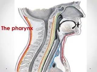
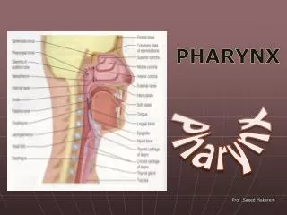
The Pharynx. Fibromuscular tube Extent Base of skull Cricoid cartilage (C6) Open anteriorly. Regions. Nasopharynx Oropharynx Laryngopharyx Systems Respiratory Digestive. Relationships. Anterior: Nasal cavity Oral cavity Larynx Posterior: Retropharyngeal space. Relationships.

The Pharynx
E N D
Presentation Transcript
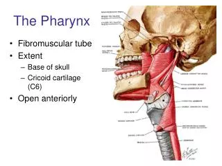
The Pharynx • Fibromuscular tube • Extent • Base of skull • Cricoid cartilage (C6) • Open anteriorly
Regions • Nasopharynx • Oropharynx • Laryngopharyx • Systems • Respiratory • Digestive
Relationships Anterior: • Nasal cavity • Oral cavity • Larynx Posterior: • Retropharyngeal space
Relationships Lateral: Carotid sheath • Internal carotid a. • Internal jugular v. • Vagus nerve (CN X) CN’s IX, XI, XII Sympathetic trunk Mandible Grant’s Atlas
Layers • Mucosa • Submucosa • Pharyngobasilar membrane/fascia • Muscularis • Adventitia • Buccopharyngeal fascia Netter
Constrictors • Superior • Middle • Inferior • Insertion: Pharyngeal raphe • Action: constrict during swallowing Grant’s Atlas
ConstrictorOrigins • Superior • Pterygo-mandibular raphe • Middle • Hyoid bone • Inferior • Thyroid and cricoid cartilages (Cricopharyngeus) S M I Grant’s Atlas
Longitudinal Muscles • Stylopharyngeus • Palatopharyngeus • Salpingopharyngeus • Action:elevate pharynx (and larynx) during swallowing SaP SC PP StP MC IC
Gaps • Gap 1 • Pharyngobasilar membrane • Gap 2 • Gap 3 • Thyrohyoid membrane • Gap 4
Gap 1 • Auditory (Eustachian, pharyngotympanic) tube • Levator palatini Grant’s Atlas
Gap 2 • Stylopharyngeus • Glossopharyngeal nerve (CN IX)
Gap 3 • Internal laryngeal nerve • Superior laryngeal artery
Gap 4 • Recurrent laryngeal nerve • Inferior laryngeal artery RL
Internal Pharynx • Nasopharynx • Oropharynx • Laryngopharynx
Nasopharynx • Posterior choanae • Auditory (Eustachian, pharyngotympanic) tube opening • Torus tubarius • Salpingopharyngeal fold • Pharyngeal recess • Pharyngeal tonsils (adenoids)
Oropharynx • Soft palate • Posterior tongue • Fauces • Palatoglossal arch • Palatopharyngeal arch • Palatine tonsil • Waldeyer’s ring
Palatine Arches Liebgott 7-79
Laryngo-pharynx • Epiglottis • Laryngeal inlet • Cricoid cartilage • Piriform recess/fossa E P C
Innervation Pharyngeal plexus • CN IX • CN XI via X • Sympathetics Motor (GSE) • CN IX to Stylopharyngeus • CN XI via X to all other muscles Grant’s Atlas
General Sensory Innervation • V2 • CN IX • CN X • Gag Reflex Clemente
Blood Supply • Ascending palatine a. (branch of facial a.) • Ascending pharyngeal a. • Superior thyroid a. • Inferior thyroid a.
VenousDrainage • Pharyngeal plexus • Drains to internal jugular vein
VenousDrainage • Pharyngeal plexus of veins • Drains to internal jugular vein • Communication with pterygoid plexus
Swallowing/Deglutition Stage 1 (Voluntary) • Jaws closed • Lips, cheeks compressed • Tongue raised Liebgott
Swallowing/Deglutition Stage 2 (Involuntary) • Nasopharynx closed off by soft palate • Larynx raised • Stylopharyngeus • Palatopharyngeus • Suprahyoids
Swallowing/Deglutition Stage 3 (Involuntary) • Larynx closed • Contraction of the three constrictors • Superior • Middle • Inferior
Swallowing/Deglutition Stage 4 (Involuntary) • Cricopharyngeus relaxes • Bolus enters esophagus • Peristalsis • Airway reopened
Dissection • Identify and separate retropharyngeal space as high as possible
Dissection • Retropharyngeal space • Disarticulate head at atlanto-occipital joint
Dissection • Prevertebral Region • Muscles • Sympathetic trunk
Be careful to • Preserve carotid sheath structures and cranial nerves • Internal jugular vein • Internal carotid a. • CN’s IX, X, XI and XII Grant’s Atlas