The Pharynx and Esophagus in Digestive Process
Learn about the anatomy and functions of the pharynx, oropharynx, laryngopharynx, and esophagus in the digestive system. Explore the passage of food, fluids, and air, and the process of deglutition. Understand the structures and mechanisms involved in the digestive processes.

The Pharynx and Esophagus in Digestive Process
E N D
Presentation Transcript
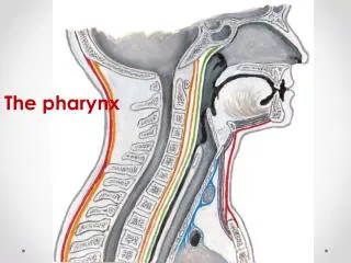
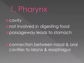
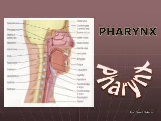
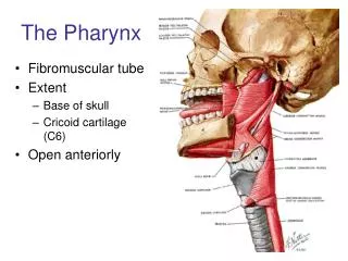
Pharynx • Oropharynx and laryngopharynx • Allow passage of food, fluids, and air • Skeletal muscle layers: inner longitudinal, outer pharyngeal constrictors
Esophagus • Flat muscular tube from laryngopharynx to stomach • Travels through the diaphragm via an opening called the esophageal hiatus • Joins stomach at the cardiac orifice
Esophagus • Esophageal glands secrete mucus to lubricate and aid in bolus movement • Muscularis: consists of skeletal muscle superiorly; smooth muscle inferiorly • Contains upper and lower esophageal sphincters: upper and lower that act as “gateways” for food • Lower esophageal sphincter prevents backflow of stomach contents into the esophagus
Mucosa (contains a stratified squamous epithelium) Submucosa (areolar connective tissue) Lumen Muscularis externa • Longitudinal layer • Circular layer Adventitia (fibrous connective tissue) (a) Figure 23.12a
Digestive Processes: Mouth • Ingestion • Mechanical digestion • Mastication is partly voluntary, partly reflexive • Chemical digestion (salivary amylase and lingual lipase) • Propulsion • Deglutition (swallowing)
Deglutition – 2 major phases • Involves the tongue, soft palate, pharynx, esophagus, and 22 muscle groups • Buccal (oral) phase • Voluntary contraction of the tongue • Pharyngeal-esophageal phase • Involuntary (sensory receptors initiate the swallowing reflex) • Control center in the medulla and lower pons
Bolus of food Tongue Pharynx Epiglottis Glottis Trachea 1 Upper esophageal sphincter is contracted. Duringthe buccal phase, the tongue presses against the hardpalate, forcing the food bolus into the oropharynxwhere the involuntary phase begins. Figure 23.13, step 1
Uvula Bolus Epiglottis Esophagus 2 The uvula and larynx rise to prevent food fromentering respiratory passageways. The tongue blocksoff the mouth. The upper esophageal sphincterrelaxes, allowing food to enter the esophagus. Figure 23.13, step 2
Bolus 3 The constrictor muscles of the pharynx contract,forcing food into the esophagus inferiorly. The upperesophageal sphincter contracts (closes) after entry. Figure 23.13, step 3
Relaxed muscles 4 Food is moved throughthe esophagus to thestomach by peristalsis. Circular musclescontract Bolus of food Longitudinal musclescontract Gastroesophagealsphincter closed Stomach Figure 23.13, step 4
5 The gastroesophagealsphincter opens, and foodenters the stomach. Relaxedmuscles Gastroesophagealsphincter opens Figure 23.13, step 5
Bolus of food Tongue Uvula Pharynx Bolus Epiglottis Epiglottis Glottis Trachea Bolus Esophagus 1 2 3 Upper esophageal sphincter iscontracted. During the buccal phase, thetongue presses against the hard palate,forcing the food bolus into the oropharynxwhere the involuntary phase begins. The uvula and larynx rise to prevent foodfrom entering respiratory passageways. Thetongue blocks off the mouth. The upperesophageal sphincter relaxes, allowing foodto enter the esophagus. The constrictor muscles of thepharynx contract, forcing foodinto the esophagus inferiorly. Theupper esophageal sphinctercontracts (closes) after entry. Relaxed muscles 4 5 Food is movedthrough the esophagusto the stomach byperistalsis. The gastroesophagealsphincter opens, and foodenters the stomach. Relaxedmuscles Circular musclescontract Bolus of food Longitudinal musclescontract Gastroesophagealsphincter closed Gastroesophagealsphincter opens Stomach Figure 23.13
The Stomach Four Main Functions: • Temporary storage for ingested food • Mechanical breakdown of food • Chemical breakdown of food • Production of intrinsic factor, necessary for absorption of vitamin B12
Stomach: Gross Anatomy • Cardiac region (cardia) • Surrounds the cardiac orifice • Fundus • Dome-shaped region beneath the diaphragm • Body • Midportion
Stomach: Gross Anatomy • Pyloric region: antrum, pyloric canal, and pylorus • Pylorus is continuous with the duodenum through the pyloric valve (sphincter) • Greater curvature • Convex lateral surface • Lesser curvature • Concave medial surface
Cardia Fundus Esophagus Muscularis externa Serosa • Longitudinal layer • Circular layer Body • Oblique layer Lumen Lesser curvature Rugae of mucosa Greater curvature Pyloric canal Pyloric antrum Duodenum Pyloric sphincter (valve) at pylorus (a) Figure 23.14a
Stomach: Gross Anatomy • Lesser omentum • From the liver to the lesser curvature • Greater omentum • Drapes from greater curvature • Anterior to the small intestine • Protects abdominal viscera
Falciform ligament Liver Gallbladder Spleen Stomach Ligamentum teres Greater omentum Small intestine Cecum (a) Figure 23.30a
Liver Gallbladder Lesser omentum Stomach Duodenum Transverse colon Small intestine Cecum Urinary bladder (b) Figure 23.30b
Stomach: Microscopic Anatomy • Still has four tunics • Muscularis and mucosa are modified • Muscularis externa • Three layers of smooth muscle • Inner oblique layer allows stomach to churn, mix, move, and physically break down food
3 muscular layers • Longitudinal • Circular • Oblique Figure 23.15a
Stomach: Microscopic Anatomy • Mucosa • Simple columnar epithelium composed of mucous cells • Produce a layer of mucus that traps bicarbonate-rich fluid beneath it • Protects the epithelial cells from acids, enzymes and abrasive materials • Gastric pits lead into gastric glands
Gastric pits Surface epithelium (mucous cells) Gastric pit Mucous neck cells Parietal cell Chief cell Gastric gland Enteroendocrine cell (b) Enlarged view of gastric pits and gastric glands Figure 23.15b
Gastric Glands • Cell types • Mucous neck cells (secrete thin, acidic mucus) • Parietal cells • Chief cells • Enteroendocrine cells
Pepsinogen Pepsin HCl Mitochondria Parietal cell Chief cell Enteroendocrine cell (c) Location of the HCl-producing parietal cells and pepsin-secreting chief cells in a gastric gland Figure 23.15c
Gastric Gland Secretions • Glands in the fundus and body produce most of the gastric juice Parietal cell secretions • HCl • pH 1.5–3.5 denatures protein in food, activates pepsin, and kills many bacteria • Intrinsic factor • Glycoprotein required for absorption of vitamin B12 in small intestine
Gastric Gland Secretions • Chief cell secretions • Inactive enzyme pepsinogen • Converted to pepsin by HCl and by pepsin itself
Homeostatic Imbalance • Gastritis: inflammation caused by anything that breaches the mucosal barrier • Peptic or gastric ulcers: erosion of the stomach wall • Most are caused by Helicobacter pylori bacteria • Cause 80% of gastric ulcers • Treated successfully with antibiotics
Bacteria Mucosa layer of stomach (b) H. pylori bacteria (a) A gastric ulcer lesion Figure 23.16
Digestive Processes in the Stomach • Physical digestion • Denaturation (breakdown) of proteins • Enzymatic digestion of proteins by pepsin (and rennin in infants) • Secretion of intrinsic factor required for absorption of vitamin B12 • Lack of intrinsic factor pernicious anemia • Delivers chyme to the small intestine
Regulation of Gastric Secretion • Regulated by CNS and hormonal mechanisms • Events occur in three phases: • Cephalic (reflex) phase: few minutes before food entry: • sight, smell, taste or thought of food initiates gastric secretion • Prepares the stomach to receive food
Regulation of Gastric Secretion 2.Gastric phase: lasts for 3–4 hours after food enters the stomach (distending stomach) • Stimulates stretch receptors • Gastrin is released increasing gastric secretion production • Distension and gastrin increase force of contraction • Intestinal phase: brief stimulatory effect as partially digested food enters the duodenum, followed by inhibitory effects to slow gastric activity down giving the intestine time to do its job
Pyloric valve closed Pyloric valve closed Pyloric valve slightly opened Propulsion: Peristaltic waves move from the fundus toward the pylorus. Grinding: The most vigorous peristalsis and mixing action occur close to the pylorus. Retropulsion: The pyloric end of the stomach acts as a pump that delivers small amounts of chyme into the duodenum, simultaneously forcing most of its contained material backward into the stomach. 1 2 3 Figure 23.19
Regulation of Gastric Emptying • As chyme enters the duodenum in 3 ml spurts • Receptors respond to stretch and chemical signals • Enterogastric reflex and enterogastrones inhibit gastric secretion and duodenal filling • Carbohydrate-rich chyme moves quickly through the duodenum • Fatty chyme remains in the duodenum 6hours or more
Small Intestine: Gross Anatomy • Major organ of digestion and absorption • 2–4 m (7-13 ft) long; from pyloric sphincter to ileocecal valve • Three Subdivisions • Duodenum – contains the bile duct and main pancreatic duct • Jejunum • Ileum
Parotid gland Mouth (oral cavity) Sublingual gland Salivary glands Tongue Submandibular gland Pharynx Esophagus Stomach Pancreas (Spleen) Liver Gallbladder Transverse colon Duodenum Descending colon Jejunum Small intestine Ascending colon Large intestine Ileum Cecum Sigmoid colon Rectum Vermiform appendix Anal canal Anus Figure 23.1
Right and left hepatic ducts of liver Cystic duct Common hepatic duct Bile duct and sphincter Accessory pancreatic duct Mucosa with folds Tail of pancreas Pancreas Gallbladder Jejunum Major duodenal papilla Main pancreatic duct and sphincter Hepatopancreatic ampulla and sphincter Duodenum Head of pancreas Figure 23.21
Structural Modifications • Increase surface area of proximal part for nutrient absorption • Circular folds (plicae circulares) • Villi • Microvilli • Circular folds • Permanent ridged • Force chyme to slowly spiral through lumen
Vein carrying blood to hepatic portal vessel Muscle layers Lumen Circular folds Villi (a) Figure 23.22a
Structural Modifications • Villi • Motile fingerlike extensions of the mucosa • Villus epithelium • Simple columnar absorptive cells (enterocytes) • Goblet cells
Structural Modifications • Microvilli • Projections (brush border) of absorptive cells • Contain brush border enzymes which complete carbohydrate and protein digestion in the small intestine
Microvilli (brush border) Absorptive cells Lacteal Goblet cell Vilus Blood capillaries Mucosa associated lymphoid tissue Enteroendocrine cells Intestinal crypt Venule Muscularis mucosae Lymphatic vessel Duodenal gland Submucosa (b) Figure 23.22b
Intestinal Juice • Secreted in response to distension or irritation of the mucosa • Slightly alkaline and isotonic with blood plasma • Largely water, enzyme-poor, but contains mucus • Facilitates transport and absorption of nutrients
Liver • Largest gland in the body • Four lobes—right, left, caudate, and quadrate
Liver • Falciform ligament • Separates the (larger) right and (smaller) left lobes • Suspends liver from the diaphragm and anterior abdominal wall • Round ligament (ligamentum teres) • Remnant of fetal umbilical vein along free edge of falciform ligament
Sternum Bare area Nipple Falciform ligament Liver Left lobe of liver Right lobe of liver Round ligament (ligamentum teres) Gallbladder (a) Figure 23.24a
Sternum Nipple Liver Bare area Lesser omentum (in fissure) Caudate lobe of liver Left lobe of liver Sulcus for inferior vena cava Porta hepatis containing hepatic artery (left) and hepatic portal vein (right) Hepatic vein (cut) Bile duct (cut) Right lobe of liver Quadrate lobe of liver Gallbladder Ligamentum teres (b) Figure 23.24b
Liver: Associated Structures • Lesser omentum anchors liver to stomach • Hepatic artery and hepatic portal vein • Bile ducts • Common hepatic duct leaves the liver • Cystic duct connects to gallbladder • Bile duct formed by the union of the above two ducts
Right and left hepatic ducts of liver Cystic duct Common hepatic duct Bile duct and sphincter Accessory pancreatic duct Mucosa with folds Tail of pancreas Pancreas Gallbladder Jejunum Major duodenal papilla Main pancreatic duct and sphincter Hepatopancreatic ampulla and sphincter Duodenum Head of pancreas Figure 23.21
Liver: Microscopic Anatomy • Liver lobules • Hexagonal structural and functional units • Filter and process blood • Composed of hepatocytes (liver cells) • Longitudinal central vein