Download
1 / 32
330 likes | 482 Vues
Explore the intricate structures and functions of the pharynx, a vital pathway for air and food. Learn about its skeletal framework, histological layers, muscular walls, and neurovascular supply.

E N D
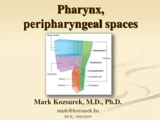
Pharynx, peripharyngealspaces Mark Kozsurek, M.D., Ph.D. mark@kozsurek.hu ED II., 18/02/2019
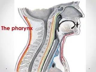
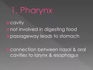
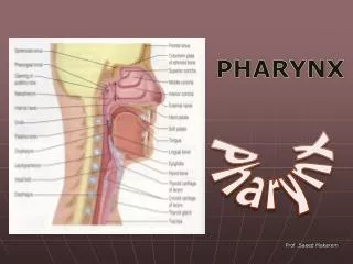
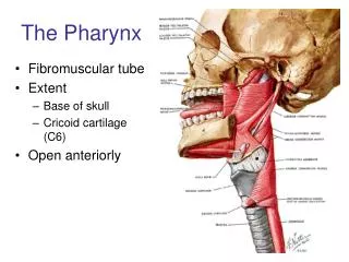
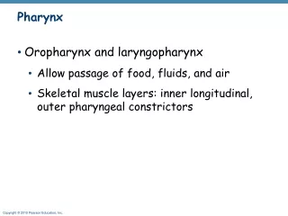
What is thepharynx? Pharynx is a musculofascial half-cylinder lacking the anterior wall. According to the anterior relations, pharynx can be devided into three parts: nasopharynx, oropharynx and laryngopharynx. It is the common pathway for the air and the food. Extends from the base of skul to the level of the 5th-6th vertebrae where is replaced by the esophagus.
Skeletalframework Anterior and superior margin of the pharynx is attached to bones, cartilages and ligaments. The two sides merge posteriorly in the midline constituting the pharyngeal raphe. Line of attachment of the pharynx to the base of the skull.
The superior line of attachment includes the posterior margin of the medial plate of the pterygoid process, the pterygoid hamulus and the pharyngomandibular raphe. The middle line of attachment follows the inferior portion of the stylohyoid ligament, the lesser horn and then the greater horn of the hyoid bone. The inferior line of attachment is represented by the oblique line of the thyroid cartilage, a tendinous thickening of the fascia of the cricothyroid muscle and the cricoid cartilage.
Pharyngealwall • Pharyngealwall is mainlyformedbyskeletalmuscles and fasciae. The latteronesareespeciallyimportantwherethemuscularwall is interrupted. • Tunicamucosa • epithelium • laminapropria • Tunicafibrosa = pharyngobasillarfascia: a thicklayerrightunderthemucosathatcoverstheinnersurface of themuscles. • Tunicamuscularis: • circularlayer: constrictors • longitudinallayer: levators • Tunicaadventitia = buccopharyngealfascia: muchthinner and is theposteriorcontinuation of thefasciacoveringtheoutersurface of thebuccinatormuscle. Histologicallymight be consideredasthetunicaadventitia of thepharynx.
Superior constrictor: pterygopharyngeal part buccopharyngeal part mylopharyngeal part glossopharyngeal part Middle constrictor: ceratopharyngeal part chondropharyngeal part Inferior constrictor: thyropharyngeal part cricopharyngeal part Note the gaps among the constrictors!!!
Levators: Salpingopharyngeus Palatopharyngeus Stylopharyngeus
Wherethemusclesaremissing... 1. At the Mylohyoid-Middle constrictor-Superior constrictor triangle: • Hyoglossus and Styloglossus muscles (Hyoglossus isolates lateral and medial sulci of the tongue) • lingual and hypoglossal nerves and deep lingual vein, submandibular duct – lateral sulcus • lingual artery and dorsal lingual vein, glossopharyngeal nerve – medial sulcus
2. At the thyrohyoid membrane: internal branch of superior laryngeal nerve (ex vagus n.), superior laryngeal artery (ex sup. thyroid a. ex ext. carotid a.), superior laryngeal vein (to sup. thyroid v. to internal jugular v.) 3. At the inferior edge of the Inferior constrictor: recurrent laryngeal nerve (ex vagus n.), inferior laryngeal artery (ex inf. thyroid a., ex thyrocervical trunk ex subclavian a.), inferior laryngeal vein (to inf. thyroid v. to left brachiocephalic v.)
Pharyngealcompartments: nasopharynx, oropharynx and laryngopharynx Identify the following structures: choana, pharyngobasillar fascia, pharyngeal fornix, pharyngeal tonsil,salpingopharyngeal fold, pharyngeal recess, salpingopalatine fold, torus tubarius, tubal tonsil, torus levatorius!
Following the removal of the mucous membrane pharyngeal muscles as well as the cartilagineous part of the tympanopharyngeal (or auditory) tube become visible.
The following structures should be identified: root of the tongue, palatoglossal and palatopharyngeal folds, tonsillary fossa, palatine tonsil, oropharynx, pharyngeal recess.
Please find: piriform recess, aryepiglottic fold, cuneiform tubercle, corniculate tubercle, interarythenoid notch, fold of the superior laryngeal nerve, greater horn of the hyoid bone, superior horn of the thyroid cartilage.
SUPERIORLY: • ascendingpharyngeal a. • (ex ext. carotid a.) • ascendingpalatine a. • (ex facial a.) • smallbranches of thelingual and maxillary a. • INFERIORLY: • pharyngealbranches of theinferiorthyroidartery • (ex thyrocervicaltrunkexsubclavian a.)
The pharyngealveinousplexuscommunicateswiththepterygoidplexus in theinfratemporal fossa thatdrainstowardthefacial and retromandibularveins and intotheexternal and internal jugularveins. • Lymphaticvesselsfromthepharynxdrainintotheretropharyngeal and paratrachealnodeswhicharesubgroups of deepcervicalnodes (situatedmainlyaroundthe internal jugularvein).
Three CNs are enough for the complete innervation of the pharynx.
Pharyngeal plexus: pharyngeal branches of the glossopharyngeal and vagus nerves and postganglionic sympathetic fibres from the superior cervical ganglion. Ambiguus nucleus (BM), dorsal vagus nucleus (GVM), lateral nucleus of ala cinerea (GVS)
In a broader sense retropharyngeal space is found behind the posterior wall of the pharynx and anterior to the prevertebral muscles. In more precise anatomical descriptions retropharyngeal space is defined as an area behind the buccopharyngeal fascia (adventitia of the pharynx) and in front of the alar fascia. As the alar fascia fuses with the pharyngeal raphe, infections can not easily spread to the contralateral side. The danger space is between the alar fascia and the prevertebral fascia and has no septum in the midline.