Download
1 / 21
240 likes | 543 Vues
White Blood Cell Differential Count. Objectives. To able to identify the different types of leucocytes under the microscope To practice the procedure for differential leucocytes counting To know normal values for the different white cell count

E N D
Objectives To able to identify the different types of leucocytes under the microscope To practice the procedure for differential leucocytes counting To know normal values for the different white cell count To know the importance of DLC in the diagnosis of disease processes.
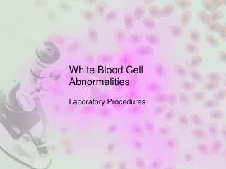
Definition The relative percentage of each type of white blood cells in peripheral blood. This experiment is a part of blood routine test.
Microscopic exam • 10× (low fold): overall smear quality, agglutination or parasites • 100× (oil Len): WBC Diff, RBC morphology
Observing direction: Observe one field and record the number of WBC according to the different type then turn to another field in the snake-liked direction *avoid repeat or miss some cells
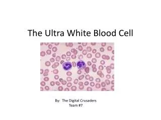
White blood cells • There are three types of granulocyte named according to their staining characteristics in blood films. They are neutrophils,eosinophils and basophils. • Agranularcells are divided into lymphocytes and monocytes.
62% of total WBCs Diameter: 10-12 microns Cytoplasm: very pale blue Granules: staining (purple) Nucleus: dark purple blue 2-5 lobes Neutrophils
Neutrophils . • neutrophilic because they owe their colour to uptake of both the acidic and the basic components of the stain
Diameter: 13-14 microns Cytoplasm : full of orange-red coarse granules Nucleus: blue 2 lobes like a pair of glass 2.3% Eosinophil
Diameter: 14-15 Cytoplasm: Granules: dark blue –black obscure nucleus Nucleus: blue 0.4% Basophil
Diameter: small 7-9 large 12-16 Cytoplasm : rim, clear, pale blue Nucleus: purple red ,oval indented 30% Lymphocyte
Normal lymphocytes • Lymphocytes are the smallest WBC. • They have large condensed nucleus, with a scanty pale blue cytoplasm.
Diameter: 16-18(are the largest normal blood cells) Cytoplasm: The cytoplasm is abundant, sky blue in colour. Some have vacuoles in the cytoplasm. Nucleus: purple, large kidney shaped and slightly indented. 5.8% Monocyte