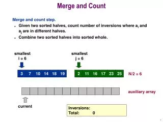
Cell count and WBC differential Count
210 likes | 1.57k Vues
Cell count and WBC differential Count. Learning objectives. After completing this laboratory exercise you should be able to: Count cells with haemocytometer and make cell suspensions. Differentiate between different types of White blood cells. Total WBC count.

Cell count and WBC differential Count
E N D
Presentation Transcript
Learning objectives After completing this laboratory exercise you should be able to: • Count cells with haemocytometer and make cell suspensions. • Differentiate between different types of White blood cells.
Total WBC count • The objective of measuring WBC count is to introduce you to the cells and tissues you will encounter in immunology. Various diseases and medical treatments alter the relative numbers of immune cells in the blood. The determination of the relative number of various leukocytes (white blood cells) in the blood is a valuable diagnostic tool in medicine and research. Because different leukocytes have different functions, changes in their proportions reveal information about the type of infection. However, knowing the proportion of these cells does not tell you about the concentration of a cell type in the blood. To get an idea of the amount or quantity, as opposed to the proportion, of various blood leukocytes, a haemocytometer is used. • A haemocytometer is a chamber of defined size that contains a grid to aid in counting numbers of cells. This count allows you to convert the umber of cells found in the chamber to the number of cells per ml of blood.
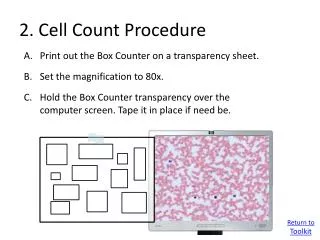
Methodology • Mix 50 µl of fresh human blood with 950 µl of 2% acetic acid solution to dilute the white blood cells and lyses all cytoplasmic membrane, . Mix gently for about 30 seconds. Be sure the cells are thoroughly suspended. • With the cover slip over the haemocytometer, fill the chamber with about 10 µl of the suspension. The fluid will be drawn into the chamber by capillary action. Allow the cells to settle on the microscope for about five minutes. Using low power (40x) or high-dry power (100x), observe and count the white blood cells in the “four corners” of the grid. A,B,C and D as in the picture below.
Methodology - Counting • Examine the setup of the haemocytometer under low power. Within the large middle square are 25 medium squares, Within each of these squares are 16 small squares. Count all the white cells lying within large four corners squares A-D each subdivided into 16 medium squares. Include those cells touching the upper and right lines. The white cells touching the left and bottom lines are not to be counted.
Calculations • To get the WBCs count in 1 ml blood you need to know the dilution factor and the volume correction factor: • The dilution factor = total volume of diluting solution / sample volume = 950 / 50 = 20 • Volume correction factor = 1 cu mm / total volume of the four large Squares • Total volume of the four large squares = Volume × Number of large squares • = (Width × Length × depth) × 4 • = (1 mm ×1 mm × 1/10 mm) × 4 = 0.4 cu mm • The volume correction factor =1 / 0.4 = 2.5 • If there are n cells in 0.4 cu mm diluted blood, then the number of cells in 1cu mm • Blood = n × dilution factor × volume correction factor • = n × 20 × 2.5 • = n × 50 • Example: • If total number of WBCs counted = 120. • Then the number of WBCs in 1 cu mm blood =120 × 50 = 6,000. • Then the number of WBCs in 1 ml blood =6000 ×1000 =6,000,000.
Differential WBC Count using Hema 3 Stain • White Blood Cells (WBCs) can be differentiated according to their staining prosperity, the differential white blood cell count determine the percentage of each type of white blood cell in a person's blood.
Preparation of Blood Smears 1. Place a small drop of diluted blood on a microscope slide as shown on this picture. 2. Slide another microscope slide toward the drop until the edge of the slide makes contact with the drop and the blood spreads out evenly along the edge of the slide (wedge smear technique). 3. As soon as this contact is made, gently pull the second microscope slide over the surface of the first to spread the blood evenly across the slide. This must be done with the right amount of speed and pressure to get a monolayer of blood cells.
Staining Method 1. Prepare the whole blood smear and allow it to completely air dry. 2. Dip smear in fixative for 5 seconds. Then, dip in Solution I for 4 seconds and then repeat the same in Solution II for 3 seconds. 3. Rinse immediately in distilled water for 20 seconds. 4. Allow the smear to air dry completely. You may wipe away the fluid from the underside of the glass to speed this process. Do not blow. 5. Examine the slide under the microscope, possibly using oil immersion. See pictures next for help in identification of cell types. 6. Make sure you can identify the various cell types. If you find a good view, show your colleagues. 7. Make a count of the cell types on the slide to get a relative proportion of the cells in the blood.
Reporting: differential WBCs • Count 100 White Blood cells in consecutive oil immersion fields. Record the number and the type of WBCs seen as a percentage. Example: Neutrophil 60% Eosinophil 4% Basophil 1% Lymphocyte 30% Monocyte 5% ________________________ total 100