Download
1 / 18
190 likes | 836 Vues
GROSS ANATOMY OF THE SPINAL CORD. Dr. Andrea D. Székely. LOCATION AND MAJOR FEATURES. The spinal cord lies in the vertebral canal and continues in the medulla oblongata. ( least modified and most caudal portion of the neural tube) SHAPE : elongated, tubular, conical

E N D
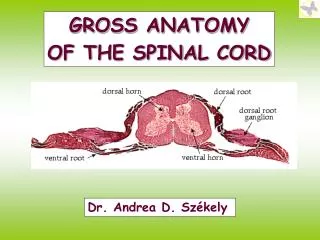
GROSS ANATOMY OF THE SPINAL CORD Dr. Andrea D. Székely
LOCATION AND MAJOR FEATURES The spinal cord lies in the vertebral canal and continues in the medulla oblongata. (least modified and most caudal portion of the neural tube) SHAPE: elongated, tubular, conical ROSTRAL END: medulla oblongata/foramen magnum CAUDAL END: conus terminalis/lumbothoracic transition ANCHORED: filum terminale (pia mater) to sacral canal/coccyx RELATIVE POSITION: fetal - corresponding to the vertebral level, newborn - conus terminalis at L3, adult - L1/L2 The spinal cord is divided into cervical, thoracic, lumbar, sacral and coccygeal regions. The grey matter is enlarged at the origins of the brachial and lumbosacral plexus (intumescentia cervicalis et lumbalis, C5-T1 and L2-L3).
EMBRYOLOGY The spinal cord develops from the NEURAL PLATE from above the notocord. With the folding of the NEURAL TUBE the centralcanal is formed (lining: ependyme). Neuroepithelial cells will give rise to neurons and glia. (Head mesenchyme) the DRG and the sympathetic neurones derive from the neural crest. The proneurones form a mantle zone (grey matter) with the axons growing towards the surface and so is the marginal zone formed (turns later into white matter). The notochord secrets a factor known as Sonic hedgehog or SHH. As a result, the floor plate then also begins to secrete SHH, and this will induce the basal plate to develop motoneurons. Meanwhile, the overlying ectoderm secretes bone morphogenetic protein (BMP). This induces the roof plate to begin to secrete BMP, which will induce the alar plate to develop sensory neurons.The alar plate and the basal plate are separated by the sulcus limitans. Additionally, the floor plate also secretes netrins. The netrins act as chemoattractants to decussation of pain and temperature sensory neurons in the alar plate across the anterior white commissure, where they then ascend towards the thalamus. The marginal zone develops into a massive passage of axans divided into 3 funiculi by the grey matter, posterior, lateral andanterior.
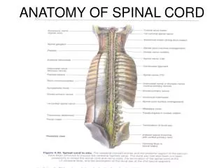
EXTERNAL FEATURES, SPINAL NERVES Anterior median fissure Posterior median sulcus (or septum) Posterior intermediate septum (only C1-C8, upper T) Lateral anterior area (ventral roots of spinal nerves) Lateral posterior sulcus (dorsal roots of spinal nerves) The spinal regions are further subdivided into external segments giving rise to pairs of nerves (C1-C8, T1-T12, L1-L5, S1-S5, Co1). The idea of segmentation is based on the pattern of nerve fibres conjoining in one spinal nerve. 31 pairs of spinal nerves arise/ pass through the intervertebral foramina. The lower lumbar and sacral nerves form the cauda equina. Nerves are composed by a dorsal (sensory) and a ventral (motor) root, except for C1 where the dorsal root may be absent in some people. The dorsal root is intersected by the accumulation of pseudounipolar sensory nerve cells (dorsal root ganglion, DRG).
THE SPINAL SEGMENT External segments Origins of nerve pairs (C1-C8, T1-T12, L1-L5, S1-S5, Co1). Basic rule : One spinal nerve is formed by all the axons emerging from one single spinal segment 1 NERVE – 1 SEGMENT Each segment is a „functional unit”, representing one body region limited independence – controlled by the brain stem and cortex) via descending tracts Intersegmental coordination - ascending fibres to higher centres - propriospinal fibres within the cord
MEMBRANES OF THE SPINAL CORD DURA MATER Envelops the cord, descends to S2 vertebra, between L2 and S2 the sac contains only the cauda equina. Each nerve passes through the intervertebral foramen retaining its dural cover, it continues as perineurium. Endorachis - connective tissue „outer layer” of the spinal dura mater - epidural space (content: fat, internal vertebral venous plexus) ARACHNOID MATER Lines the dural cavity, rather large subarachnoideal, but insignificant subdural space. LUMBAR PUNCTION - below L2 PIA MATER Adheres to the spinal cord and roots, its lateral aspect gives rise to the denticulate ligament (21 on each side) reaching the arachnoid mater. It lies between the dorsal and ventral nerve roots.
THE CLINICAL ANATOMY OF SPINAL ANESTHESIA LAYERS TO PENETRATE Skin Supraspinal ligament Interspinal ligament Lig. flavum Endorachis Epidural space Dura mater spinalis Arachnoid mater Subarachnoid space
VASCULAR SUPPLY OF THE SPINAL CORD ARTERIES 2 anterior spinal arteries (later fuse) supply the grey matter, and parts of the lateral and anterior funiculus. 2 posterior spinal arteries supply the dorsal horns and the dorsal funiculus. Arcuate anastomoses exist between the sides Longitudinal vascular system: contain the spinal branches (from the vertebral, intercostal, lumbar, lateral sacral vessels and supply the vertebrae, meninges, the nerve roots and the medulla, joining the anterior and posterior spinal branches. Radicular system: vessels following the nerve roots supply the spinal segments. Radicular branches arise segmentally from the ascending cervical, deep cervical, intercostal, lumbar and sacral arteries. Anterior radicular branches: 6-10, left predominance. Largest branch: artery of Adamkiewicz - apparent atthe lumbar enlargement. Posterior radicular branches: 10-23, no explicit left predominance. Vulnerable segments: T1-T3 posteriorly, T3 and L1 anteriorly
VASCULAR SUPPLY OF THE SPINAL CORD VEINS (NO VALVES) The pattern of vessels is similar to that of the arteries. Anterior longitudinal venous trunk 1 anteromedian vein: takes up the sulcal veins 2 anterolateral veins: take up the ant-lat vessels Posterior longitudinal venous trunk 1 posteromedian vein: drains the posterior funiculus 2 posterolateral veins: drain the dorsal horns+ a strip of the lateral funiculus. The vessels drain into 5-10 posterior radicular veins VENOUS VASOCORONA - coronal anastomotic veins connecting the longitudinal vessels The blood is drained into the internal vertebral venous plexus (epidural venous plexus), where the plexus is connected to thoracic, abdominal and intercostal veins, then to the external vertebral venous plexus to reach the azygos and hemiazygos veins. N.B. connections to the prostatic plexus represent a route by which malignous tumors may metastasize!!
DERMATOMES dermatome a piece of skin innervated by the same spinal nerve C 8, Th 12, L 5 und S 5 At the edes there are always overlapping supply fields
CLINICAL RELEVANCES WHAT MAY DAMAGE THE SPINAL CORD? - Pressure (e.g. tumor or discus hernia), - Rupture (horizontal damage separating the segments causing paralysis), - Inflammations (Myelitis, multiplex sclerosis), - Failing blood supply - Degenerative procedures DISCUS HERNIA
CROSS SECTIONS OF THE SPINAL CORD CERVICAL LEVEL
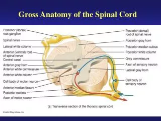
CROSS SECTIONS OF THE SPINAL CORD THORACIC LEVEL
CROSS SECTIONS OF THE SPINAL CORD LUMBAR LEVEL
CROSS SECTIONS OF THE SPINAL CORD SACRAL LEVEL