Digestion Lab
Digestion Lab. Experiment #1: Carbohydrate Digestion. Tube 1 3 ml water Tube 2 3 ml 0.2% amylase Tube 3 3 ml 0.2% amylase + 10 drops of 1.0M HCl Tube 4 3 ml 0.2% amylase place in hot water bath for 5 min. 4. 2. 3. 1. 4. 4. 4. 2. 2. 2. 3. 3. 3. 1. 1. 1.

Digestion Lab
E N D
Presentation Transcript
Experiment #1:Carbohydrate Digestion • Tube 1 • 3 ml water • Tube 2 • 3 ml 0.2% amylase • Tube 3 • 3 ml 0.2% amylase + 10 drops of 1.0M HCl • Tube 4 • 3 ml 0.2% amylase • place in hot water bath for 5 min 4 2 3 1
4 4 4 2 2 2 3 3 3 1 1 1 Experiment #1:Carbohydrate Digestion • Add 5.0 ml starch solution to each tube • Incubate in 37C bath for 1.5 hr • Divide contents of each tube evenly into 2 tubes • Lugol’s Test • Benedict’s Test
4 2 3 1 Experiment #1:Carbohydrate Digestion • Lugol’s Test • presence of starch • add a few drops of Lugol's reagent (iodine) • if starch absent, transparent brown color • if starch present, opaque black-blue color or
4 2 3 1 Experiment #1:Carbohydrate Digestion • Benedict’s Test • presence of maltose • add 5.0 ml of Benedict's reagent • immerse in hot water bath for 2 min • rate the amt of maltose present • blue = - (none) • green = + (a little bit) • yellow = ++ (some) • orange = +++ (lots)
5 2 3 1 Experiment #2:Protein Digestion • Add egg white into each tube • Tube 1 • 10 drops of water + 5.0 ml pepsin • Tube 2 • 10 drops of 1M HCl + 5.0 ml pepsin • Tube 3 • 10 drops of 1M HCl + 5.0 ml pepsin • place in ice bath • Tube 4 • 10 drops of 1M HCl + 5.0 ml water • Tube 5 • 10 drops of 1M NaOH + 5.0 ml pepsin 4
Experiment #2:Protein Digestion • Incubate tubes 1,2,4 and 5 at 37 C for 1.5 hours • Observe any digestion of egg white undigested digested
Experiment #3:Fat Digestion • add 3.0 ml of cream to each tube • Tube 1 • 5.0 ml water + few grains of bile salts • Tube 2 • 5.0 ml pancreatin • Tube 3 • 5.0 ml pancreatin + few grains of bile salts
Experiment #3:Fat Digestion • Test pH of each solution w/ pH probe • rinse probe w/ detergent after each test • Place in 37C bath • Retest pH at 20, 40 and 60 min
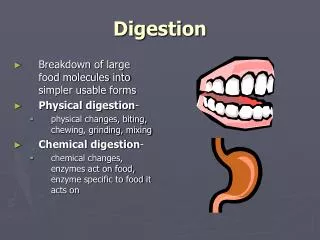
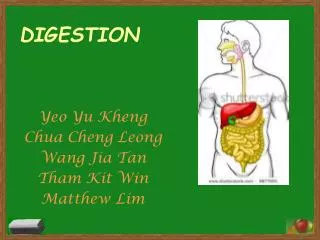
Digestion • Physical and chemical break down nutrients into absorbable unit • Physical digestion (chewing, mixing) • Chemical digestion (enzyme catalyzed) • polysaccharides monosaccharides • proteins amino acids • fats glycerol + fatty acids
Enzymes Protein Catalysts • speed up the rate of chemical reactions • are not permanently altered in the reactions • do not change the nature of the reaction
Factors Affecting Enzyme Activity • Temperature • Temp, kinetic energy, reaction rate • high Temp changes structure of enzymes • ’s enzyme function Lecture Fig 4.3
Factors Affecting Enzyme Activity • pH • 3D structure of enzymes changes at different pH • optimal enzyme function at specific pH • function at higher or lower pH’s Lecture Fig 4.4
Carbohydrate Digestion • begins with salivary amylase (ptyalin) • breaks starch (polysaccharide) into maltose (disaccharide) • Simple sugars = reducing sugars • Drive reduction reactions for other substances • Become oxidized Lecture Fig 18.1
Oxidation-Reduction Reactions • oxidation reaction • reaction in which a molecule loses e-s • reduction reaction • reaction in which a molecule gains e-s • Example • NADH → NAD+ = oxidation • NAD+ → NADH = reduction • O2 + 4H+ + 4e- → 2H2O = reduction
Benedict’s Test • Cu2+ + Maltose (reduced) → Cu+ + Maltose (oxidized) • 4 Cu+ + O2 → Cu2O (orange color)
Protein Digestion • Begins in the stomach • Gastric Epithelial Cells • Parietal Cells • Secrete HCl • Chief Cells • Secrete Pepsinogen • Low pH activates pepsinogen • Pepsinogen autocatalyzes self into pepsin • Cleaves proteins Lecture Figs 18.1, 18.9
Protein Digestion • Continues in small intestine • chyme enters pyloric sphincter • intestine releases hormone (secretin), that stimulates the release of pancreatic juices • Chymotrypsin, trypsin, etc. • Enzymes activated in intestine • Digest small polypeptides into amino acids Lecture Figs 18.26, 18.29
Fat Digestion • Begins in stomach • Most in small intestine • (pancreatic and intestinal lipases) • Fats are nonpolar! • Digestion depends on the presence of bile from the gall bladder • emulsification breaks up fat into small droplets • lipases break triglycerides into monoglycerides + fatty acids • form micelles in intestinal lumen Lecture Figs 18.1, 18.35, 18.36
Fat Digestion • absorbed by the epithelium • reform triglycerides in the epithelial cells • combined w/ protein to form chylomicrons which are secreted into lacteals • carried via lymph to the veins Lecture Figs 18.1, 18.35, 18.36
Fat Digestion • Triglycerides → glycerol + fatty acids • Fatty acids lower pH of aqueous solutions • ↑ fat digestion, ↓pH Lecture Fig 18.1