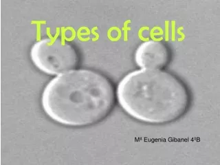
Types of Cells--Nerve, Muscle, Immune, and Skin
Types of Cells--Nerve, Muscle, Immune, and Skin. Nerve Cells. The human body is made up of trillions of cells. Cells of the nervous system, called nerve cells or neurons , are specialized to carry “messages” through an electrochemical process.

Types of Cells--Nerve, Muscle, Immune, and Skin
E N D
Presentation Transcript
Nerve Cells The human body is made up of trillions of cells. Cells of the nervous system, called nerve cells or neurons, are specialized to carry “messages” through an electrochemical process. The human brain has approximately 100 billion neurons. Neurons come in many different shapes and sizes. Some of the smallest neurons have cell bodies that are only 4 microns wide. Some of the biggest neurons have cell bodies that are 100 microns wide (Remember, 1 micron is equal to one thousandth of a millimeter!).
Nerve Cells Neurons are similar to other cells in the body because: • Neurons are surrounded by a cell membrane. • Neurons have a nucleus that contains genes. • Neurons contain cytoplasm, mitochondria and other organelles. • Neurons carry out basic cellular processes such as protein synthesis and energy production.
Nerve Cells However, neurons differ from other cells in the body because: • Neurons have specialized cell parts called dendrites and axons. Dendrites bring electrical signals TO the cell body and axons take information AWAY from the cell body. • Neurons communicate with each other through an electrochemical process. • Neurons contain some specialized structures (synapses) and chemicals (neurotransmitters)
Connection Point to Previous Knowledge: Exocytosis and Neurotransmitters • When nerve cells release large molecules, called neurotransmitters (a chemical), they are releasing them through exocytosis. This is the “chemical” part of electrochemical communication between neurons. • Remember, what does exocytosis require? (aka what kind of transport is it?) https://www.youtube.com/watch?v=WhowH0kb7n0
Nerve Cells What is inside of a neuron? A neuron has many of the same organelles such as mitochondria, cytoplasm and a nucleus, as other cells in the body. • Nucleus - contains genetic material (chromosomes) including information for cell development and synthesis of proteins necessary for cell maintenance and survival. Covered by a membrane. • Nucleolus - produces ribosomes necessary for translation of genetic information into proteins • Nissl Bodies - groups of ribosomes used for protein synthesis. • Endoplasmic reticulum (ER) - system of tubes for transport of materials within cytoplasm. Can have ribosomes (rough ER) or no ribosomes (smooth ER). With ribosomes, the ER is important for protein synthesis. • Golgi Apparatus - membrane-bound structure important in packaging peptides and proteins (including neurotransmitters) into vesicles. • Microfilaments/Neurotubules - system of transport for materials within a neuron and may be used for structural support. • Mitochondria - produce energy to fuel cellular activities.
Nerve Cells Neurons can be classified by the direction that they send information. • Sensory neurons: send information from sensory receptors (e.g. in skin, eyes, nose, tongue, ears) TOWARD the central nervous system. • Motor neurons: send information AWAY from the central nervous system to muscles or glands. • Interneurons: send information BETWEEN sensory neurons and motor neurons. Most interneurons are located in the central nervous system.
Connection Point to Previous Knowledge: The Famous Sodium Potassium Pump! • What type of transport is this? Does it require energy (ATP)? • Which ions are transported across the membrane? Nerve cells send electrical signals through their axon to communicate information from one cell to the next. This is the “electrical” part of the electrochemical communication along the neuron and is called action potential. https://www.youtube.com/watch?v=b2ctEsGEpe0
Nerve Cells--Review Video: https://www.youtube.com/watch?v=FT44xI92LCU&t=23s (2:41 min)
Muscle Cells Bones don’t work alone – they need help from the muscles and joints. Muscles pull on the joints, allowing us to move. They also help your body perform other functions so you can grow and remain strong, such as chewing food and then moving it through the digestive system. The human body has more than 650 muscles, which make up half of a person’s body weight. They are connected to bones by tough, cord-like tissues called tendons, which allow the muscles to pull on bones. If you wiggle your fingers, you can see the tendons on the back of your hand move as they do their work.
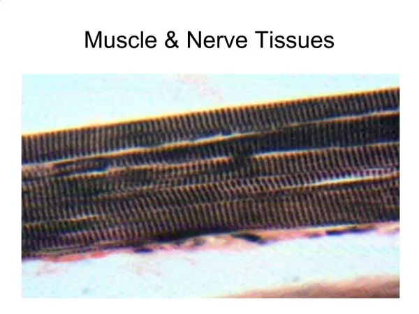
Muscle Cells Humans have three different kinds of muscle: 1. Skeletal muscle is attached to bone, mostly in the legs, arms, and abdomen, chest, neck and face. • Skeletal muscles are called striated because they are made up of fibers that have horizontal stripes when viewed under a microscope. • These muscles hold the skeleton together, give the body shape, and help it with everyday movements (known as voluntary muscles because you can control their movement). • They can contract (shorten or tighten) quickly and powerfully, but they tire easily and have to rest between workouts.
Muscle Cells 2. Smooth, or involuntary, muscle is also made of fibers, but this type of muscle looks smooth, not striated. • Generally, we can’t consciously control our smooth muscles; rather, they’re controlled by the nervous system automatically (which is why they’re also called involuntary). • Examples of smooth muscles are the walls of the stomach and intestines, which help break up food and move it through the digestive system. • Smooth muscle is also found in the walls of blood vessels, where it squeezes the stream of blood flowing through the vessels to help maintain blood pressure. • Smooth muscles take longer to contract than skeletal muscles do, but they can stay contracted for a long time because they don’t tire easily.
Muscle Cells 3. Cardiac muscle is found in the heart. • The walls of the heart’s chambers are composed almost entirely of cardiac muscle fibers. • Cardiac muscle is also an involuntary type of muscle. • Its rhythmic, powerful contractions force blood out of the heart as it beats.
Muscle Cells • What do you notice about the similarities between the types of muscle cells? • What are the main differences between each of the muscle cells?
Muscle Cells Our Nonstop Muscles Even when we sit perfectly still, muscles throughout the body are constantly moving. Muscles enable the heart to beat, the chest to rise and fall during breathing, and blood vessels to help regulate the pressure and flow of blood through the body. When we smile and talk, muscles help us to communicate, and when we exercise, they help us to stay physically fit and healthy. The movements your muscles make are coordinated by the brain and nervous system. The involuntary muscles are controlled by structures deep within the brain and upper part of the spinal cord called the brain stem. The voluntary muscles are regulated by the parts of the brain known as the cerebral motor cortex and the cerebellum.
Muscle Cells When you decide to move, the motor cortex sends an electrical signal through the spinal cord and peripheral nerves to the muscles, causing them to contract. The motor cortex on the right side of the brain controls the muscles on the left side of the body and vice versa. The cerebellum coordinates the muscle movements ordered by the motor cortex. Sensors in the muscles and joints send messages back through peripheral nerves to tell the cerebellum and other parts of the brain where and how the arm or leg is moving and what position it’s in. This feedback results in smooth, coordinated motion. If you want to lift your arm, your brain sends a message to the muscles in your arm and you move it. When you run, the messages to the brain are more involved, because many muscles have to work in rhythm.
Muscle Cells Muscles move body parts by contracting and then relaxing. Muscles can pull bones, but they can’t push them back to the original position. So they work in pairs of flexors and extensors. The flexor contracts to bend a limb at a joint. Then, when the movement is completed, the flexor relaxes and the extensor contracts to extend or straighten the limb at the same joint. For example, the biceps muscle, in the front of the upper arm, is a flexor and the triceps, at the back of the upper arm, is an extensor. When you bend at your elbow, the biceps contracts and then the biceps relaxes and the triceps contracts to straighten the elbow.
Skeletal Muscle Contraction: M Lines and Z Lines • Skeletal muscle fibers are composed of many myofibrils. • Myofibrils contain units called sarcomeres. • Each sarcomere consists of alternating thick and thin protein filaments, giving skeletal muscles its striated appearance. The muscle contracts when these filaments slide past each other. • The thick filaments are myosin, which are anchored at the center of the sarcomere, called the M Line. • The thin filaments are composed of actin, which are anchored to the Z lines on the outer edges of the sarcomere.
Skeletal Muscle Contraction: M Lines and Z Lines When a skeletal muscle contracts: • The sarcomere shortens from both sides when actin filaments slide along the myosin filaments. The myosin filament actually pulls the actin along its length, using ATP for energy. • The myosin head extends and attaches to binding sites on the actin, forming a cross-bridge. • The myosin pulls the actin filament toward the M line, thereby shortening the sarcomere.
Skeletal Muscle Contraction: M Lines and Z Lines • Muscle contractions are controlled by the actions of calcium. • The thin actin filaments have associated proteins called troponin and tropomyosin, which allow the muscle to contract or relax. • When calcium levels are high enough and ATP is present, calcium ions bind to the troponin, which displaces tropomyosin, exposing the myosin binding sites on actin. This allows myosin to attach to actin, forming a cross-bridge and causing the muscle to contract. • When a muscle is relaxed, tropomyosin blocks the cross-bridge binding sites on actin (so that the myosin cannot attach).
Muscle Cells Review Skeletal: https://edpuzzle.com/media/59fa0dac70192b40e1e97d99 Smooth: https://www.youtube.com/watch?v=yzQAgfivX74 Cardiac: https://www.youtube.com/watch?v=gBcdtqnQnbo Muscle Contraction: https://www.youtube.com/watch?v=ousflrOzQHc
Immune Cells What Are Immune Cells? An army is a force of people fighting together. The immune system is just like that, except it's a collection of cells fighting together. The immune cells are the soldiers that defend our bodies from invaders. Just like different soldiers have different jobs, so do different immune cells. The immune system has two parts: innate and adaptive. Different immune cells play roles in each of them.
Immune Cells The innate immune system is very general and includes the skin, mucus, and saliva. It also contains cells that travel around the body looking for things that are suspicious. Immune cells have receptors that allow them to detect certain things (like bacterial cell proteins and dsRNA) that are unique to bacteria, viruses, or fungi. In other words, they recognize non-human things. Some immune cells find these things and then get rid of them by eating them! They ingest the sketchy invader in a process called phagocytosis and then break it down (part of endocytosis). One type of phagocytic cell is called a neutrophil. Neutrophils hang out in the blood and can get called to particular sites of infection. Another type of phagocytic cell is a macrophage. Macrophages can move here and there around the body, but they really like to hang out in the spleen and lymph nodes where they see a lot of action.
Immune Cells • Natural Killer Cells are just what they sound like: tough, assassin-type cells. These guys look for problems within our own cells. If it looks like a cell has been hijacked by a virus or cancer, the natural killers move into action. However, they don't eat their prey. Instead, they release chemicals to get the job done. • Eosinophils are similar to Natural Killer Cells because they don't engulf their prey. These cells hang out near mucus membranes and look for things like parasites. They release enzymes to destroy any invaders. • Dendritic Cells are a link between the innate and the adaptive immune system. They are sentinels that look for fishy things near the skin. If they find something, they eat it, but then bring it to a lymph node and deliver it to the adaptive immune system.
Immune Cells Unlike the innate immune system, the adaptive immune system relies on fewer types of cells to carry out its tasks: B cells and T cells. The cells of the adaptive immune system are lymphocytes. B cells, which are derived from the bone marrow, become the cells that produce antibodies. T cells, which mature in the thymus, differentiate into cells that either participate in lymphocyte maturation, or kill virus-infected cells.
Immune Cells After T cells develop in the thymus, they either travel around in the blood or lymphatic system or migrate to different organs in the body. As soon as a specific invader stimulates them, helper T cells produce chemicals. Some chemicals trigger B cells to develop into plasma cells, while others stimulate killer T cells to target and kill cells that may have either become infected by the invader or are cancerous. Regulatory T cells help to control the immune reaction to prevent it getting out of hand. Natural killer T cells also produce chemicals to help regulate the immune response and protect against invaders and tumors. Memory T cells stay around for a long time after the immune response has finished. In this way, they can react quickly if the same invader appears again and multiply to produce a large number of T cells to eliminate it.
Immune Cells • Unlike T cells, B cells cannot directly attack infected cells. Instead, B cells primarily produce proteins called antibodies that can hijack invaders as they travel in the blood. • When they come across invaders, B cells are stimulated into action and produce plasma cells and memory B cells. Each plasma cell is specialized to make a particular antibody -- a specialized protein to attack a specific invader. • The function of antibodies is to act as a flag on the infected cell so T cells recognize which cells to destroy. When invaders become coated with antibody, they are more easily targeted by other proteins in the immune system, as well as by the specialized cells known as phagocytes that are responsible for eating foreign substances and infected cells. • While plasma cells disappear after an immune response is finished, memory B cells stay around for a long time. If the same invader appears again, antibodies are already available to help fight it off.
How Diseases Spread Direct contact: An easy way to catch most infectious diseases is by coming in contact with a person or animal who has the infection. Three ways infectious diseases can be spread through direct contact are: • Person to person. A common way for infectious diseases to spread is through the direct transfer of bacteria, viruses or other germs from one person to another. This can occur when an individual with the bacterium or virus touches, kisses, or coughs or sneezes on someone who isn't infected. These germs can also spread through the exchange of body fluids from sexual contact. The person who passes the germ may have no symptoms of the disease, but may simply be a carrier.
How Diseases Spread • Animal to person. Being bitten or scratched by an infected animal — even a pet — can make you sick and, in extreme circumstances, can be fatal. Handling animal waste can be hazardous, too. For example, you can acquire a toxoplasmosis infection by scooping your cat's litter box. • Mother to unborn child. A pregnant woman may pass germs that cause infectious diseases to her unborn baby. Some germs can pass through the placenta. Germs in the vagina can be transmitted to the baby during birth.
How Diseases Spread Indirect contact: Disease-causing organisms also can be passed by indirect contact. Many germs can linger on an inanimate object, such as a tabletop, doorknob or faucet handle. When you touch a doorknob handled by someone ill with the flu or a cold, for example, you can pick up the germs he or she left behind. If you then touch your eyes, mouth or nose before washing your hands, you may become infected. • Insect bites Some germs rely on insect carriers — such as mosquitoes, fleas, lice or ticks — to move from host to host. These carriers are known as vectors. Mosquitoes can carry the malaria parasite or West Nile virus, and deer ticks may carry the bacterium that causes Lyme disease. • Food contamination Another way disease-causing germs can infect you is through contaminated food and water. This mechanism of transmission allows germs to be spread to many people through a single source. E. coli, for example, is a bacterium present in or on certain foods — such as undercooked hamburger or unpasteurized fruit juice.
Immune Cells Video: https://www.youtube.com/watch?v=HNP1EAYLhOs (3:01 min)
Skin Cells The skin is the body’s largest organ. It serves many important functions, including regulating body temperature, maintaining water and electrolyte balance, and sensing painful and pleasant stimuli. The skin keeps vital chemicals and nutrients in the body while providing a barrier against dangerous substances from entering the body and provides a shield from the harmful effects of ultraviolet radiation emitted by the sun. In addition, skin color, texture, and folds, help mark people as individuals. Anything that interferes with skin function or causes changes in appearance can have important consequences for physical and mental health.
Skin Cells The skin has three layers that each performs specific tasks: • Epidermis • Dermis • Fat layer (also called the subcutaneous layer)
Skin Cells The epidermis is the relatively thin, tough, outer layer of the skin. Most of the cells in the epidermis are keratinocytes. They originate from cells in the deepest layer of the epidermis called the basal layer. New keratinocytes slowly migrate up toward the surface of the epidermis. Once the keratinocytes reach the skin surface, they are gradually shed and are replaced by newer cells pushed up from below. The outermost portion of the epidermis, known as the stratum corneum, is relatively waterproof and, when undamaged, prevents most bacteria, viruses, and other foreign substances from entering the body. The epidermis also protects the internal organs, muscles, nerves, and blood vessels against trauma. In certain areas of the body that require greater protections (such as the palms of the hands and the soles of the feet), the outer keratin layer of the epidermis (stratum corneum) is much thicker.
Skin Cells Scattered throughout the basal layer of the epidermis are cells called melanocytes, which produce the pigment melanin, one of the main contributors to skin color. Melanin’s primary function, however, is to filter out ultraviolet radiation from sunlight, which damages DNA, resulting in numerous harmful effects, including skin cancer. The epidermis also contains Langerhans cells, which are part of the skin’s immune system. Although these cells help detect foreign substances and defend the body against infection, they also play a role in the development of skin allergies.
Skin Cells The dermis, the skin’s next layer, is a thick layer of fibrous and elastic tissue (made mostly of collagen, elastin, and fibrillin) that gives the skin its flexibility and strength. The dermis contains nerve endings, sweat glands and oil (sebaceous) glands, hair follicles, and blood vessels. The nerve endings sense pain, touch, pressure, and temperature. Some areas of the skin contain more nerve endings than others. For example, the fingertips and toes contain many nerves and are extremely sensitive to touch. The sweat glands produce sweat in response to heat and stress. Sweat is composed of water, salt, and other chemicals. As sweat evaporates off the skin, it helps cool the body. Specialized sweat glands in the armpits and the genital region (apocrine sweat glands) secrete a thick, oily sweat that produces a characteristic body odor when the sweat is digested by the skin bacteria in those areas.
Skin Cells The sebaceous glands secrete sebum into hair follicles. Sebum is an oil that keeps the skin moist and soft and acts as a barrier against foreign substances. The hair follicles produce the various types of hair found throughout the body. Hair not only contributes to a person’s appearance but has a number of important physical roles, including regulating body temperature, providing protection from injury, and enhancing sensation. A portion of the follicle also contains stem cells capable of re-growing damaged epidermis. The blood vessels of the dermis provide nutrients to the skin and help regulate body temperature. Heat makes the blood vessels enlarge (dilate), allowing large amounts of blood to circulate near the skin surface, where the heat can be released. Cold makes the blood vessels narrow (constrict), retaining the body’s heat. Over different parts of the body, the number of nerve endings, sweat glands and sebaceous glands, hair follicles, and blood vessels varies. The top of the head, for example, has many hair follicles, whereas the soles of the feet have none.
Skin Cells Below the dermis lies a layer of fat that helps insulate the body from heat and cold, provides protective padding, and serves as an energy storage area. The fat is contained in living cells, called fat cells, held together by fibrous tissue. The fat layer varies in thickness, from a fraction of an inch on the eyelids to several inches on the abdomen and buttocks in some people.
Fight or Flight Response https://learn.genetics.utah.edu/content/cells/cellcom/ • How do skin cells play a role in the “fight or flight” response? Which parts of the skin cells are activated by signal molecules during the response?
Fight or Flight Response When our fight or flight response is activated, sequences of nerve cell firing occur and chemicals like adrenaline, noradrenaline and cortisol are released into our bloodstream. These patterns of nerve cell firing and chemical release cause our body to undergo a series of very dramatic changes: Our respiratory rate increases. Blood is shunted away from our digestive tract and directed into our muscles and limbs, which require extra energy and fuel for running and fighting. Our pupils dilate. Our awareness intensifies. Our sight sharpens. Our impulses quicken. Our perception of pain diminishes. Our immune system mobilizes with increased activation. We become prepared—physically and psychologically—for fight or flight. We scan and search our environment, "looking for the enemy."
Fight or Flight Response Some of the physical signs that may indicate that the fight-or-flight response has kicked in include: • Rapid Heartbeat and Breathing: The body increases heartbeat and respiration rate in order to provide the energy and oxygen to the body that will be needed to fuel a rapid response to the danger. • Pale or Flushed Skin: As the stress response starts to take hold, blood flow to the surface areas of the body is reduced and flow to the muscles, brain, legs, and arms are increased. You might become pale as a result, or your face may alternate between pale and flushed as blood rushes to your head and brain. The body's blood clotting ability also increases in order to prevent excess blood loss in the event of injury. • Dilated Pupils: The body also prepares itself to be more aware and observant of the surroundings during times of danger. Another common symptom of the fight-or-flight response is the dilation of the pupils, which allows more light into the eyes and results in better vision of the surroundings. • Trembling: In the face of stress or danger, your muscles become tense and primed for action. This tension can result in trembling or shaking.
Fight or Flight Response-Skin’s Response • In the skin, epinephrine (adrenaline) binds to a receptor on an erector pilli smooth muscle cell. • This causes a signaling cascade that contracts the muscle, raising the hair on the surface of the skin. • On the surface of sweat glands, epinephrine binds to Alpha-1 adrenergic receptors, triggering a signaling cascade that contracts the gland, squeezing sweat to the skin’s surface.