Download

1 / 30
340 likes | 803 Vues
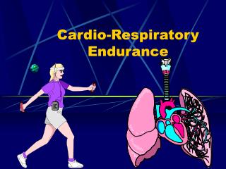
Impaired aerobic capacity/endurance. Min H. Huang, PT, PhD, NCS. Learning Objectives. Describe causes and factors contributing to impaired aerobic capacity in older adults.

E N D
Impaired aerobic capacity/endurance Min H. Huang, PT, PhD, NCS
Learning Objectives • Describe causes and factors contributing to impaired aerobic capacity in older adults. • Describes physical therapist patient management to address decreased endurance and its impact on function for a geriatric client.
Reading assignments • Guccione: Ch 12(pp.229-233, Box 12-2, Box 12-3 only)
Cardiovascular functional changes with age • Decreases • Cardiac Output: resting and maximal • Max HR • Resting and maximal stroke volume • Venous Return • Blood Flow • 25% increase in Left Ventricular thickness • Vessel rigidity • 65 yo has 30-40% of aerobic capacity of young adult • Increases • Blood Pressure: both resting & exercise BP • Cardiac Dysrhythmias • By 75 yo, <10% of SA node cells
Age related changes in the heart • Increase adipose tissue • Increase collagen content • Decrease muscle cells (myocytes) • Increase cardiomyocyte senescence • Decrease innervation/nerve conduction tissue • Decrease sympathetic modulation of HR Results in decrease excitability, decrease cardiac output, venous return and an INCREASE in dysrhythmias.
Age-dependent changes to cardiovascular tissues North B J , and Sinclair D A Circulation Research 2012;110:1097-1108
Age related changes in the heart • With the walls of the heart becoming less compliant • Declines in Left ventricle expansion and contractility (i.e. reduced end diastole volume) • Results in decreased ejection fraction (Frank-Starling law) • Increased atrial size correlates to Left ventricular compliance • increased workload on the atria • hypertrophy of the aorta
Ventricular function curves showing the Frank–Starling relationship Hanft2008. http://cardiovascres.oxfordjournals.org/content/77/4/627.full.pdf+html
Cardiac hypertrophy • A reduction in cardiac output due to aging stimulates the myocardium to compensate by increasing muscle mass • short-term enhancement of cardiac output • long-term impact on cardiac function • Ventricular hypertrophy results from an increase in size of individual cardiomyocytes • physiological and reversible, e.g. exercise-induced • pathological and irreversible, i.e. disease-based
Valvular changes with aging • Age related valvular circumference • Mitral and Aortic valves have the most issues. • Thickening and calcification of the cusps and leaflets • Lose water content. • Results in valvular stenosis and mitral valve insufficiency: heart murmurs J. Blackwood
Aging of the peripheral vasculature • Arterial thickening and stiffness as well as dysfunctional endothelium • Increase systolic pressure, increased risks of atherosclerosis, HTN, stroke, A-fib, ischemia • Decrease in elasticity of vessel walls may result in chronic rigidity or vessel wall diameter • In venous system: valves become stiff and incompetent Impaired return of blood, may play a role in phlebitis and thrombus formation
Aging changes in cardiac conduction • Declines in function and number of pacemaker cells in SA node • By age 70 only 10% of the number found in young adults are present • Proliferation of fibrous tissue in nerve conduction system may affect SA function • Incidence of Sick Sinus Syndrome (SSS) increases with age • Bradycardia, SA node arrest, SA exit conduction block
Age related changes on ECG • ~50% of older adults have cardiac conduction abnormalities at rest. • PR and QT intervals have small increases • ST segment becomes flattened • Amplitude of the T wave diminishes
Oxygen consumption (V·O2) • A physiological measure of how much oxygen the body uses at rest or during activity • Increases in proportion to intensity of exercise/physical activity and will plateau when maximal ability for oxygen delivery is reached, which is maximal oxygen consumption (V·O2 max). • Maximal oxygen consumption is directly related to aerobic capacity
Heart rate response to an aerobic exercise bout and adaptation following aerobic exercise training
Stroke volume response to an aerobic exercise bout and adaptation following aerobic exercise training
Cardiac output response to an aerobic exercise bout and adaptation following aerobic exercise training
Arteriovenous oxygen difference (a-vO2diff) response to an aerobic exercise bout and adaptation following aerobic exercise training.
Warning signs during PT • SBP > 180 mmHg and/or DBP > 100 mmHg at rest • HR >100 bpm at rest (consider HRmax or PRE > 13 with exercise ) • Excessive dyspnea • Low angina threshold • Claudication pain =DVT • Lack of HR or BP response with activity or excessive response with activity • Drop of SBP >20mmHg or HR > 10 bpm with exercise • Slow recovery from activity (>3-5 min)
STOP PT session immediately • Complaints of light-headedness, confusion, dyspnea, or onset of angina • Syncope or near syncope • Nausea • Unusual or severe fatigue • Staggering or persistent unsteadiness • Severe claudication or other pain • Angina • Abnormal HR or BP response to exercise
Aerobic and Strength training • Aerobic training allows for improved CV fitness, decrease in HTN, improves lipid metabolism, prevents Left ventricular hypertrophy • Strength training allows for improvements in overall strength, muscle mass and quality J. Blackwood
Guidelines for exercise interventions with cardiovascular • Consider: intensity, mode, frequency, duration, and progression • Monitor: HR, BP, SaO2, ECG, BORG scale (RPE), estimated VO2 max, MET levels. • Be aware of the medication side effects (orthostatic hypotension, blunted HR) that can occur with this population • Refer to ex physiology for reference; Also: AHA, AACVPR, ACSM J. Blackwood
Guidelines for exercise interventions with cardiovascular • Cardiac clients with L vent dysfunction or cardiac induced ischemia do not have improvements in max aerobic capacity, but do with submax strengthening. • Poor aerobic capacity: not able to sustain adequate HR and BP with exercise. • Make changes in an exercise program with the geriatric client in response to the CV or CP signs that occur.