Download
1 / 23
240 likes | 304 Vues
Explore the internal factors influencing the heart's function. Learn about cardiac muscle cells, signal propagation, and the cardiac cycle. Discover the importance of conducting fibers and blood pressure in heart health.

E N D
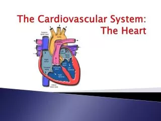
Option D: Human Physiology D.4 The heart http://i.livescience.com/images/i/000/057/264/original/human-body-heart-130925.jpg?1380117166
D.4 • Essential idea: Internal and external factors influence heart function.
D.4.U1 Structure of cardiac muscle cells allows propagation of stimuli through the heart wall. Cardiac muscle cells are and join end to end in a complex network. They are (due to the arrangement of the contractile proteins actin and myosin), they may have , and they are not under voluntary control. They also have and are which prevent them from becoming fatigued.
D.4.U1 Structure of cardiac muscle cells allows propagation of stimuli through the heart wall. Cardiac muscle cells have special structures that allow the propagation of stimuli through the heart wall: Intercalated discs – Specialized junction consisting of a double membrane containing . Gap junctions have . This allows the (ion movement) to . Branching – The y-shape allows the cells to be and also allows the from one cell to the network of cells. These adaptations allow for synchronization of muscle contraction.
12 6 1 7 11 2 8 5 9 3 10 4
D.4.U2 Signals from the sinoatrial node that cause contraction cannot pass directly from atria to ventricles. SL valves AV valves The cardiac cycle includes the sequence of events when the heart beats. The heart contracts without nerve stimulation. This is achieved by a group of specialized cells called the . Gap junctions allow the contraction to spread rapidly across the entire atrium causing the atria to undergo systole (contract). The signal sent from the .
D.4.U3 There is a delay between the passing on of a stimulus at the AV node. AND D.4.U4 This delay allows time for atrial systole before the atrioventricular valves close. SL valves AV valves To stimulate the ventricle to contract, the signal reaches another specialized group of cells called the . There is approximately a before the AV node sends out its own action potential. Why do you think there needs to be a delay before the ventricle contracts???
D.4.U5 Conducting fibers ensure coordinated contraction of the entire ventricle wall. The signal from the AV node spreads through fibers to specialized tissue called . This signal causes the ventricle to undergo . Remember, the ventricle walls are much thicker than the atria and therefore require this system of conducting fibers and Purkinje fibers.
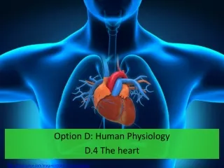
REVIEW 6.2.A2 Pressure changes in the left atrium, left ventricle and aorta during the cardiac cycle. AND D.4.U6 Normal heart sounds are caused by the atrioventricular valves and semilunar valves closing causing changes in blood flow. SL valves AV valves Atria contract Ventricles relaxed Blood pushed into ventricles Atrial pressure increases slightly higher (no big increase needed as much of the blood already moved passively to ventricles) AV valves open SL valves closed Atria relaxed Ventricles contract Blood pushed into arteries Early: Ventricular pressure increases above atrial pressure AV valves close “LUB” SL valves closed Late: Ventricular pressure increases above artery pressure SL valves open Atria and ventricles relaxed Blood flows into atria from veins Atrial pressure higher than ventricular but lower than artery pressure AV valves open SL valves closed “DUB” AV valves close “LUB” SL valves closed “DUB”
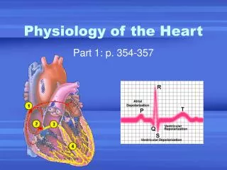
D.4.S3 Mapping of the cardiac cycle to a normal ECG trace. • An electrocardiogram (ECG) is a • It can be used to determine if electrical activity is normal and it may be used to determine if parts of the heart are too large or overworked.
D.4.S3 Mapping of the cardiac cycle to a normal ECG trace. P wave: Point Q: QRS complex: T wave: (ions returning to resting potential)
Voltage/mV Time/s http://4.bp.blogspot.com/_5Nslwo9F6bI/S_EU-Kcs4DI/AAAAAAAAAg4/5f0VSazrcN4/s1600/ECG+trace+%26+basics.jpg
The period between the points R and R’ represent one cardiac cycle R’ R Q Q S S
D.4.S1 Measurement and interpretation of the heart rate under different conditions. We addressed this skill in the labs we performed for the Blood System unit. Heart rate can be detected in a number of ways: pulse in wrist using fingers not thumb, side of neck below jaw, or use of hand-grip heart monitors like we used. Different conditions could include types of exercise, intensity of exercise, recovery from exercise, relaxation, body position including lying down, breathing and breath holding. http://www.pcrush.com/images/300/827631.jpg http://cdn1.medicalnewstoday.com/content/images/articles/258/258118/two-left-fingers-feeling-a-pulse-on-right-wrist.jpg http://images.agoramedia.com/everydayhealth/gcms/pg-sixty-second-health-checks-05-full.jpg
D.4.S2 Interpretation of systolic and diastolic blood pressure measurements. Blood Pressure and Rate of Flow • During ventricular systole, • During ventricular diastole, Blood pressure is measured to record the regular cycle of pressure in the arteries as the heart contracts • Usually measured in upper arm (brachial artery) with a sphygmomanometer and stethoscope • Average pressure for young adult male is
D.4.A1 Use of artificial pacemakers to regulate the heart rate. Common heart problems - Pacemakers In some individuals the sinoatrial node (aka pacemaker) malfunctions or they may have a block in the signal conduction pathway. “Bradycardia is a heartbeat that is slower than normal. Heart block is a disorder that occurs if an electrical signal is slowed or disrupted as it moves through the heart. Heart block can happen as a result of aging, damage to the heart from a heart attack, or other conditions that disrupt the heart's electrical activity. Some nerve and muscle disorders also can cause heart block, including muscular dystrophy.” https://www.nhlbi.nih.gov/health/health-topics/topics/pace/whoneeds
D.4.A1 Use of artificial pacemakers to regulate the heart rate. Common heart problems - Pacemakers To regulate the heartbeat, patients may be fitted with an . The device is placed under the skin in the chest area. It has wires (leads) that are threaded through a blood vessel that leads into the interior of the heart. The leads . Placement of the leads will depend upon the patient’s particular heart problem. https://www.nhlbi.nih.gov/health/health-topics/topics/pace/howdoes
D.4.A2 Use of defibrillation to treat life-threatening cardiac conditions. Common heart problems – Heart attack & Defibrillators Heart attacks (cardiac arrest) happens when the . Abnormalities, such as , in the cardiac cycle can occur. When this happens the . To restore a normal heart rhythm, first responders use a to first detect if fibrillation is occurring and if it is, an electrical discharge is given off. *An AED is a portable defibrillator. http://www.nhlbi.nih.gov/health/health-topics/topics/aed/howtouse
D.4.A3 Causes and consequences of hypertension and thrombosis. Common heart problems – Hypertension Hypertension is . This cannot be determined by one blood pressure measure as many factors can increase blood pressure, such as stress. It is best to regularly monitor your blood pressure. • Causes: . Recall that plaque formation often is the result of high levels of lipids and cholesterol in blood. • Consequences: • Damage to artery cells can lead to them becoming narrower and stiff • Weakening of artery causing an aneurysm (bulge in artery wall) that can burst and cause internal bleeding • Stroke due to blood vessels in the brain becoming weak and narrow, leak or rupture or clots in arteries • Kidney failure due to damage to arteries leading to kidneys and capillaries in the glomerulus http://blogs.furman.edu/wellness/files/2014/11/1-heart-e.jpg
D.4.A3 Causes and consequences of hypertension and thrombosis. Thrombosis is the condition when a . Common heart problems – Thrombosis One form of thrombosis is (DVT) • Causes: can cause a clot to form in one of the larger veins, usually the leg. (Slow-flowing blood is more likely to clot) • Consequences: • All or a portion of the clot can break loose and travel to a smaller vein, blocking blood flow (especially dangerous if the clot becomes lodged in a vein in the lung) http://www.stjohnprovidence.org/upload/images/Hospital%20and%20Centers/SJ%20Macomb-Oakland%20Hospital%20Macomb%20Center/Interventional%20Radiology/DVT_normal_and_embolus.gif
D.4.A3 Causes and consequences of hypertension and thrombosis. Thrombosis is the condition when a clot (thrombus) forms in a blood vessel. Common heart problems – Thrombosis Another form of thrombosis is Causes: causing substantial narrowing of the lumen resulting in slow blood flow and clot formation. • Consequences: • Heart attack
D.4.S4 Analysis of epidemiological data relating to the incidence of coronary heart disease. Epidemiology is the branch of medicine that deals with the incidence, distribution, and possible control of diseases and other factors relating to health. Coronary heart disease (CHD) refers to damage to the heart caused by a reduction in blood supply to the heart. Read pages 526-527 in your textbook to see who is at risk of CHD and how it can be prevented. http://www.world-heart-federation.org/fileadmin/user_upload/images/CVD_Health/worldmapOverview.jpg