Cell Adhesion and Junctional Structures in Epithelial Cells
550 likes | 751 Vues
Learn about the significance of cell adhesion in tissue organization and pathology, as well as the role of adhesion molecules like cadherins, selectins, and integrins in forming cellular junctions. Discover how these molecules contribute to the structure and function of epithelial cells.

Cell Adhesion and Junctional Structures in Epithelial Cells
E N D
Presentation Transcript
Cell adhesion, junctional structures, the epithelial cell Prof. Dr. Pál Röhlich Dr. Anna L. Kiss Department of Anatomy, Histology and Embryology Semmelweis University Budapest 2017
Adhesion • Specificbindingsbetweena.) individualcells • b.) cells and extracellularmatrix (ECM). • - Integratescells in a multicellularorganism, • - crucialfororganisation of tissues and organs. • - intracellularanchoringtofilaments of thecytoskeleton (actinmicrofilamentsorintermediatefilaments). • Adhesionbetweencells • Adhesionbetweencell and extracelluarmatrix
I.Adhesion between cells: • Adhesionproteinsin theplasmamembrane (integralmembraneproteins). • Significance: • inembryonicdevelopment(cellassociations, switchingbetweencellassociations, specificattachmentbetweennerve and muscle, formation of interneuronalconnections (synapses), formationofepitheliallayers, etc. …) • inadultorgnisms: • longlastingconnections (e.g. epithelia, nervoussystem) • ortemporalconnections(e.g. intheimmunesystem). • pathology: metastasis of cancercells, …
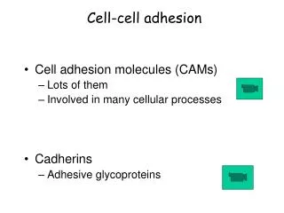
Cell Adhesion Molecules (CAMs)- are intramembrane (transmembrane)proteinslocated on the cell surface; - involved with the binding with other cells or with the extracellular matrix (ECM) in the process called celladhesion;- many weak bonds are formed between the cells. These proteins are typically transmembrane receptorsand are composed of three domains:a.) an intracellular domain that interacts with the cytoskeleton,b.) a transmembrane domain, c.) an extracellular domain that interacts either with other CAMs of the same kind (homophilicor homotypicbinding) or with other CAMs or the extracellular matrix (heterophilicor heterotypicbinding).
Celladhesionmolecules homotypic binding heterotypic binding
Celladhesionmolecules Ca2+-dependent: cadherins selectins integrins Ca2+-independent: IgG-like-CAMs (immunoglobulinsuperfamily)
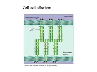
Cell-cell adhesion molecules • Ca2+-dependent: • Cadherins: Adhesionproteinswith a singletransmembraneportion. • The extracellular part usuallyconsists of 5 repeatingdomains. Extracellulartip of themolecule (lastdomain) binds a similardomain of cadherin intheoppositemembrane(homotypicbinding). • Stability of thedomains is Ca2+-dependent(withoutCa2+binding, theextracellular part of themoleculecollapses and thecellsbecomedissociated). • Cadherinsform a relativelystrongbindingbetweencells.
Cadherin-superfamily (about 180 cadherin types in human, „variations on a theme”), Nomination according to cell type and characters, recently numbers. Classical cadherins:E-cadherin (epithelial, in cells of epithelium and morula), P-cadherin (placentar, in heart, lung, gut), N-cadherin(neural, in development of the nervous system, synaptogenesis), M-cadherin (muscular, myoblasts), R-cadherin (retinal, outgrowth of nerves), VE-cadherin(vascular endothelial), K-cadherin (kidney), … In a single cell several cadherin types can occur, expression of cadherins. Appearance of E-cadherin in the morula stage of embryonic development. Mouse. Cells of the morula closely adhere to one another: compaction. 1.5 days 3.5 days
2. Selectins Ca2+- dependent, weak heterotypic bindings. The terminal domain has binding site for certain membrane glycoproteins of other cells. Role: recirculation of lymphocytes in the organism, adhesion of leucocyte to the endothelial surface of blood vessels. („rolling”), … L-Selectin (on leucocytes), P-selectin (on blood platelets), E-selectin (on endothelium) anchoring proteins actin filaments
3. Integrins: Ca2+-dependent, heterotypicbinding, α and βchains, • binding site ontheextracellular end. • 24 differentintegrins. Largemajoritybindstocomponents of theextracellularmatrix, but a few of themtocells. Theseare: LFA-1 (αLβ2) Integrin (onleucocytes), binds I-CAM and V-CAM adhesionmoleculesonendothelialcells. Migration of leucocytesthroughthewallofthebloodvessel. Mac-1 (αMβ2) Integrin (onmacrophages and lymphocytes) Binding site α-chain β-chain
Ca2+-independent Immunoglobulin-like adhesion molecules: CAMs No Ca-dependence! Immunoglobulin-domains. Weak adhesion, fine regulation. N-CAM (neural cell adhesion molecule). Homotypic bindings. Negative charges on the sialic acid molecules can modulate binding. Role: in development of the nervous system, formation of ganglia, outgrowth of nerve fibers. I-CAM, V-CAM(in the lining cells of blood vessels). Heterotypic bindings with integrins of leucocytes. L1-CAM(cell migration, differentiation), PECAM (adhesion between blood platelets and endothelium). Disulfide-bond
II. Adhesion between cells and extracellular matrix (ECM) Strong connection between cells and ECM ECM: produced by the cells: 1.) fibers; 2.) proteoglycans, glucosaminoglycans; 3.) matrix binding glycoproteins 1.) Connective tissue fibers: Collagen fibers: Large family of collagen proteins. Most frequently occur: collagen type I: composed of fibrous collagen molecules, resistant against pull, thin collagen fibrils form bundles of various thicknesses: thick collagen fibers. collagen type III: can form thin fibers often interconnected with each other (reticular fibers).
2.) Glycosaminoglycans, proteoglycans. Glycosaminoglycans (GAGs) are long sugar chains, composed of disaccharide units and carrying acidic groups (carboxyl, sulfate), therefore they contain many negative charges. High water-binding capacity! Proteoglycans (PGs):consist of a core polypeptide chain and of GAG chains laterally bound with covalent bonds.
3.) Matrix-binding glycoproteins: heterogenous proteins many binding sites forECM components and cells. They form bridges in between them. The most promeinent matrix-binding proteins: laminin és a fibronectin.
II.Adhesion between cells and extracellular matrix (ECM): integrins : Functions of integrins: • Adhesion between cell and ECM (Attachment of the cell onto the underlying structure is important!) • Local integration of the cytoskeleton with the ECM • Signal transductionfrom inside outward and reversed Indirectsignificance: cellmigration, celldivision, development, differenciation
Integrin as signal transduction molecule Active and inactive forms. Binding of a ligand to the integrin molecule on one side of the membrane activates the molecule on the other side of the membrane (transduction of a signal across the membrane). The role of talin. Activation of signal transduction pathways (binding to the β-chain activates extracellular part of the molecule which in turn strongly binds ECM components.) ligand binding talin binding
Integrins play important role in cell migration Integrin internalistion: via caveolae Recycling/or lysosomal degradation
The epithelial cell Surface epithelium: Epithelial cells are attached to each other laterally and form a continuous layer on free surfaces. General characteristics of the epithelial cell are introduced on the example of the columnar epithelium. The epithelial cell is a polarized cell: cell organelles, cytoskeletal constituents, membrane domains and cell junctions are arranged in a characteristic pattern and orientation. Apical (luminal) surface apical surface Golgi gap junction centrosome lateral surface Intestinal epithelium basal lamina basal surface Columnar epithelial cell showing localisation of cell junctions and surfaces.
Epithelialcells Poligonal cells apical lateral basal
Apical surface • glykocalyx • microvilli (brush border) • stereocilium • cilia, flagellum Function: • protection (mechanical, biological) • diffusion barrier • absorption, • movements • secretion (exocytosis) • transcytosis
Glycocalyx Function cell-cell recognition (MHC, blood groups) enzyme-function (intestine: enterocytes) cell membrane
Microvilli (brush border) Microfilaments Glykocalyx 0.08 m 1 m Terminal web – bundles of actin
Microvilli (brush border) To increase the surface
Stereocilia length: 8-10 µm diameter: 1 µm actin filaments stereocilia Ductus epidydymis
Cilia length: 5-10 µm diameter: 0,2 µm Function: movements Structure: microtubules (tubulin) nexin (connectsmicrotubules) dynein (outer and inner)
Basal surface Basement membrane: inhibits the free diffusion induces polarity regeneration filter (kidney) epithelial cell • lamina basalis lamina lucida (rara) lamina densa • lamina fibro-reticularis
Basal surface Basal or basement membrane (light microscope) • Basal lamina: TypeIV-collagen + adhesion molecules Glycoproteins (laminin, fibronectin) + proteoglycans • Lamina rara externa (lamina lucida) • Lamina densa • Lamina rara interna • Lamina fibroreticularis: TypeIII-collagen
Basement membrane A thin layer (40-120 nm) under the basal surface of the epithelium, only seen with the electron microscope lamina rara lamina densa Electron microscopy lamina fibroreticularis
Molecular structure of the basal lamina. Components: Collagen type IV(these fibrous protein molecules form a multilayered network: lamina densa, a resistant and plastic layer) Laminin(matrix-binding molecule) Perlecan(proteoglycan) Nidogen(a small protein with multiple binding sites) Fibronectin(matrix-binding molecule) Binding to the plasma membrane by: integrin (laminin-receptor), dystroglycan and syndecan (membrane proteoglycans) Perlecan Dystroglycan Integrin Laminin Nidogen lamina densa collagen Type IV Fibronectin
Biological significance of the basal lamina: • mechanical: a.) bindsthe epithelial layer firmly to the ECM. Important e.g. in the strong binding of the epidermis to the underlying connective tissue. The basal lamina is a continuous layer, b.) inhibits immigration of connective tissue cells into the epithelium or free emigration of epithelial cells into the connective tissue (cancer!). • cell biological: the basal lamina sends information signals through integrins into the cell interior and is therefore important in survival and division of the epithelial cell as well as in the maintenance of cell polarity. During epithelial regeneration it „leads” the epithelial cells to cell-free areas. Without basal lamina no continuous epithelial layer can be formed. Genetic defects of basal lamina proteins are lethal for the embryo in an early developmental stage. 3. molecular sieve: filters blood from the capillaries of the renal glomerulus (ultrafiltrate) and retains proteins and cells in the blood. In genetic defects of one of its molecular components blood or blood proteins appear in the urine. The pore size of the filter depends primarily on proteoglycans in the basal lamina.
Basal striation membrane invaginations+mitochondria
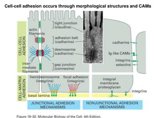
Lateralsurface: celljunctions • Mechanical connecting structures: • macula adherens (desmosoma) • zonula adherens • hemidesmosoma • Diffusion barriers • zonula occludens (tight junction) • Communicating junctions • nexus (gap junction) Long-lasting coupling between cells and between cell and ECM with various functions. General structure: adhesion molecules, adaptor proteins,cytoskeleton
Desmosomes and zonula adherens adaptor proteins: (cateins, vinculin, α-actinin) adaptor proteins integral membrane protein, adhesion mol.
Hemidesmosome (half-desmosome): Adhesion molecules: integrins (e.g. laminin receptor α6β4) Adaptor molecules: plectin, dystonin Cytoskeleton: intermediate filaments (e.g. keratin in epithelium) ECM: laminin (lamina basalis)
Hemidesmosomes • It makes stronger the connection of the epithelial cells to the lamina basalis integrins
Zonula occludens (tight junction) • Integralmembrane protein: occludin, claudin • adapter molecules: ZO1, ZO2 • cytoskeleton: microfilaments (actin)í
Zonula occludens (tight junction) ZO1, ZO2 protein
Functions of tightjunction • Diffusionbarrierintheplasmamembrane(blockinglateraldiffusioninthemembrane), thebelt-liketightjunctiondividestheplasmamembraneinto an apical and baso-lateraldomain (withdifferentsets of molecules) • Diffusionbarrierintheintercellularspace: free diffusion of substancesbetweenneighboringcells is blocked. • Biologicalsignificance:controlled and unidirectionaltransportacrosstheepitheliallayer apical membrane baso-lateral membrane baso-lateral membrane
Zonula occludens (tight junction) Cell junction functioning as a diffusion barrierin the intercellular space and in the plasma membrane. Localisation: belt-like, running circumferentially close to the luminal surface.
Nexus (gap junction) Patch-like contacts between two cells: large number of channels (connexons) in the opposite membranes, that are bound to each other in the intercellular space to form continuous channels between the two cells. cell membranes intercellular space connexon (connexin subunits) 2 connexons
Molecular structure: connexon: a complex composed of 6 transmembrane proteins(connexins), that surround a central canal. A similar complex in the opposite membrane is joined to it in the intercellular space. A continuous canal is formed which leads from one cell into the other making communication by free diffusion of low molecular weight substances possible. Connexin:Transmembrane proteins Gap junction:patch-like membrane domain with densely packed connexons Diversity of connexons, in different cell types. Combinations of different connexins in a connexon. Examples of connexin isoforms: Cx50 in the crystalline lens of th eye, defects lead to glaucoma Cx26 in sensory cells of the inner ear, defects cause auditory malfunction Cx32 in nerve fibers, defects lead to problems in nerve conduction. opposite plasma membranes
Nexus Biological significance: Communication between cells: nutrient transport (cells of the lens, osteocytes, follicular cells surrounding the oocyte in the ovary), synchronized reaction for signals during the development etc. Electrical connection between cells: electrical synapsis: ions can migrate between cells, stimulus can be transmitted without delay; In heart muscle: synchronized contraction
Nexus (gap junction, macula communicans) gap junction
Cell junctions in polarized cells Hemidesmosomes