Download
1 / 85
851 likes | 878 Vues
Learn how cells reproduce through division, passing hereditary information via DNA contained in chromosomes. Discover the structure and function of nucleotides in DNA and the process of DNA replication.

E N D
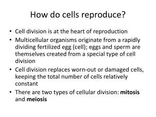
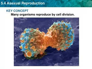
Cells reproduce by cell division. One cell gives rise to two or more cells, called daughter cells. (Virchow) Each daughter cell receives a complete set of heredity information—identical to the information in the parent cell—and about half of the cytoplasm.
Cell division transmits hereditary information to each daughter cell. The hereditary information in each cell is deoxyribonucleic acid (DNA). DNA is contained in chromosomes. A molecule of DNA consists of smaller subunits called nucleotides.
The structure of DNA phosphate nucleotide T A base A sugar C G G C C G C T A A C G A T A T T (a) (b) A single strand of DNA The double helix Fig. 8-1
The eukaryotic chromosome consists of DNA bound to protein. Human chromosomes contain a single DNA double helix that is 50 to 250 million nucleotides long, which would be about 3 inches long if the DNA were completely relaxed.
The nucleotides are held together by hydrogen bonding between the bases in two strands, called a double helix, which looks like a twisted ladder. James Watson and Francis Crick combined the X-ray data with bonding theory to deduce the structure of DNA
DNA is composed of four nucleotides DNA is made of chains of small subunits called nucleotides Each nucleotide has three components • A phosphate group • A deoxyribose sugar • One of four nitrogen-containing bases • Thymine (T) • Cytosine (C) • Adenine (A) • Guanine (G)
The structure of DNA phosphate nucleotide T A base A sugar C G G C C G C T A A C G A T A T T (a) (b) A single strand of DNA The double helix Fig. 8-1
Hydrogen bonds between complementary bases hold two DNA strands together in a double helix (continued) Because of their structures and the way they face each other, adenine (A) bonds only with thymine (T) and guanine (G) bonds only with cytosine (C) Bases that bond with each other are called complementary base pairs Thus, if one strand has the base sequence CGTTTAGCCC, the other strand must have the sequence GCAAATCGGG
Parental DNA double helix The parental DNA is unwound New DNA strands are synthesized with bases complementary to the parental strands Each new double helix is composed of one parental strand (blue) and one new strand (red)
Segments of different lengths along a DNA molecule are the units of inheritance called genes. Each gene spells out the instructions for making the proteins of the cell. When a cell divides, it first replicates its DNA, and each copy is transferred into each daughter cell.
a pair of homologous chromosomes Both chromosomes carry the same allele of the gene at this locus; the organism is homozygous at this locus gene loci This locus contains another gene for which the organism is homozygous Each chromosome carries a different allele of this gene, so the organism is heterozygous at this locus the chromosome from the male parent the chromosome from the female parent
Homologous chromosomes carry the same kinds of genes for the same characteristics Genes for the same characteristic are found at the same loci on both homologous chromosomes Genes for a characteristic found on homologous chromosomes may not be identical Alternative versions of genes found at the same gene locus are called alleles
a pair of homologous chromosomes Both chromosomes carry the same allele of the gene at this locus; the organism is homozygous at this locus gene loci This locus contains another gene for which the organism is homozygous Each chromosome carries a different allele of this gene, so the organism is heterozygous at this locus the chromosome from the male parent the chromosome from the female parent
An organism’s two alleles may be the same or different Each cell carries two alleles per characteristic, one on each of the two homologous chromosomes If both homologous chromosomes carry the same allele (gene form) at a given gene locus, the organism is homozygous at that locus If two homologous chromosomes carry different alleles at a given locus, the organism is heterozygous at that locus (a hybrid)
a pair of homologous chromosomes Both chromosomes carry the same allele of the gene at this locus; the organism is homozygous at this locus gene loci This locus contains another gene for which the organism is homozygous Each chromosome carries a different allele of this gene, so the organism is heterozygous at this locus the chromosome from the male parent the chromosome from the female parent
Unlike prokaryotic chromosomes, eukaryotic chromosomes are separated from the cytoplasm by a membrane-bound nucleus. Eukaryotic cells always have multiple chromosomes. Eukaryotic chromosomes contain more DNA than prokaryotic chromosomes.
Eukaryotic chromosomes usually occur in pairs (continued). The nonreproductive cells of many organisms have chromosomes in pairs, with both members of the pair being the same length. The chromosomes are the same length and have the same staining properties because they have the same genes arranged in the same order.
Eukaryotic chromosomes usually occur in pairs. An entire set of stained chromosomes from a single cell is called a karyotype. sexchromosomes Fig. 8-6
The number of different types of chromosomes in a species is called the haploid number and is designated n. In humans, n = 23. Diploid cells contain 2n chromosomes. Humans body cells contain 2n = 46 (2 x 23) chromosomes.
A typical human cell has 23 pairs of chromosomes. 22 of these pairs have a similar appearance and are called autosomes. Human cells also have a pair of sex chromosomes, which differ from each other in appearance and in genetic composition. Females have two X chromosomes. Males have one X and one Y chromosome.
Eukaryotic chromosomes usually occur in pairs. An entire set of stained chromosomes from a single cell is called a karyotype. sexchromosomes Fig. 8-6
There are two types of division in Eukarytic cells: mitotic cell division and meiotic cell division. Mitotic cell division may be thought of as ordinary cell division, such as occurs during development from a fertilized egg, during asexual reproduction, and in skin, liver, and the digestive tract every day. Meiotic cell division is a specialized type of cell division required for sexual reproduction.
Meiotic cell division Meiosis
Cell division is required for sexual and asexual reproduction. Sexual reproduction in eukaryotic organisms occurs when offspring are produced by the fusion of gametes (sperm and eggs) from two adults. Gametes are produced by meiotic cell division, (Meiosis which results in daughter cells with exactly half of the genetic information of their parent cells). Fertilization of an egg by a sperm results in the restoration of the full complement of hereditary information in the offspring.
Mitotic cell division Mitosis
Reproduction in which offspring are formed from a single parent, without having a sperm fertilize an egg, is called asexual reproduction. Asexual reproduction produces offspring that are genetically identical to the parent. Examples of asexual reproduction occur in bacteria, single-celled eukaryotic organisms, multicellular organisms such as Hydra, and many trees, plants, and fungi.
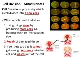
Cell division is required for growth and development. Cell division in which the daughter cells are genetically identical to the parent cell is called Mitotic cell division. After cell division, the daughter cells may grow and divide again, or may differentiate, becoming specialized for specific functions. The repeating pattern of division, growth, and differentiation followed again by division is called the cell cycle.
Duplicated chromosomes separate during cell division. Prior to cell division, the DNA within each chromosome is replicated. The duplicated chromosomes then consist of two DNA double helixes and associated proteins that are attached to each other at the centromere.
Eukaryotic chromosomes during cell division centromere genes duplicatedchromosome(2 DNA doublehelices) sisterchromatids (a) A replicated chromosome consists of two sister chromatids independentdaughterchromosomes,each with oneidentical DNA double helix (b) Sister chromatids separate during cell division Fig. 8-5
Duplicated chromosomes separate during cell division (continued). Each of the duplicated chromosomes attached at the centromere is called a sister chromatid. During mitotic cell division, the sister chromatids separate and each becomes a separate chromosome that is delivered to one of the two resulting daughter cells.
Eukaryotic chromosomes during cell division centromere genes duplicatedchromosome(2 DNA doublehelices) sisterchromatids (a) A replicated chromosome consists of two sister chromatids independentdaughterchromosomes,each with oneidentical DNA double helix (b) Sister chromatids separate during cell division Fig. 8-5
Meiotic cell division Meiosis
Cell division is required for sexual and asexual reproduction. Sexual reproduction in eukaryotic organisms occurs when offspring are produced by the fusion of gametes (sperm and eggs) from two adults. Gametes are produced by meiotic cell division, (Meiosis which results in daughter cells with exactly half of the genetic information of their parent cells). Fertilization of an egg by a sperm results in the restoration of the full complement of hereditary information in the offspring.
Mitotic cell division Mitosis
Mitotic cell division Mitotic cell division consists of nuclear division (called mitosis) followed by cytoplasmic division (called cytokinesis) and the formation of two daughter cells.
The eukaryotic cell cycle is divided into two major phases: interphase and cell division. During interphase, the cell acquires nutrients from its environment, grows, and duplicates its chromosomes. During cell division, one copy of each chromosome and half of the cytoplasm are parceled out into each of two daughter cells.
Mitosis is divided into four phases. Prophase Metaphase Anaphase Telophase
Prophase Fig. 8-9b–c
During prophase, the chromosomes condense and are captured by the spindle microtubules. Three major events happen in prophase: The duplicated chromosomes condense. The spindle microtubules form. The chromosomes are captured by the spindle.
The centriole pairs migrate with the spindle poles to opposite sides of the nucleus. When the cell divides, each daughter cell receives a centriole. Every sister chromatid has a structure called a kinetochore located at the centromere, which attaches to a spindle apparatus.
Prophase Fig. 8-9b–c
Metaphase Fig. 8-9d
During metaphase, the chromosomes line up along the equator of the cell. At this phase, the spindle apparatus lines up the sister chromatids at the equator, with one kinetochore facing each cell pole.
Metaphase Fig. 8-9d
Anaphase Fig. 8-9e
During anaphase, sister chromatids separate and move to opposite poles of the cell. Sister chromatids separate, becoming independent daughter chromosomes. The kinetochores pull the chromosomes poleward along the spindle microtubules.
Anaphase Fig. 8-9e
Telophase Fig. 8-9f