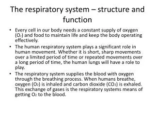
Structure of the Respiratory system
210 likes | 508 Vues
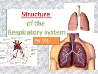
Structure of the Respiratory system. P6 M3 . Respiratory System: Intakes oxygen Releases carbon dioxide waste Circulatory system: Transports gases in blood between lungs and cells. Respiratory system works with the Cardiovascular system….

Structure of the Respiratory system
E N D
Presentation Transcript
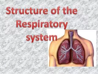
Structure of the Respiratory system P6 M3
Respiratory System: Intakes oxygen Releases carbon dioxide waste Circulatory system: Transports gases in blood between lungs and cells Respiratory system works with the Cardiovascular system…
Task… In order to gain a head start on the assessment you guys need to find a blank pictures of the various respiratory system structures and begin to label them: nasal cavity epiglottis pharynx larynx trachea bronchus bronchioles lungs (lobes, pleural membrane, thoracic cavity, visceral pleura, pleural fluid, alveoli) diaphragm intercostal muscles (external and internal)
The Importance of the RS • If respiratory system and/or circulatory system fails, death will occur • Cells need O2 for work; release CO2 as a waste product • Accumulation of excess CO2 is toxic to cells and MUST be removed
Respiratory Structures and Organs: Explained • Nasal cavity • Space above and behind the nose • Made of cartilage and bone • Divided into 2 by a cartilaginous septum • Hairs within the nostrils filter out dust etc before air passes into two nasal cavities. • Designed to warm, moisten, and filter air before it passes to the nasopharynx • A mucous layer • Pharynx – (throat) • Funnel shaped • Connects to larynx and oesophagus • Small muscular tube • conducts food and air • exchanges air with Eustachian tube to equalize pressure
Respiratory Structures and Organs: Explained • Epiglottis • flap of cartilage that covers trachea • ensures food travels down the esophagus • Larynx – (voice box) • Connects the pharynx and the trachea. • Made of cartilage and muscle • contains vocal cords • Helps us to speak
larynx trachea bronchi bronchioles Respiratory Structures and Organs: Explained • Trachea – (windpipe) • Tubular passageway (12 cm long 2 cm diameter) to carry air towards the lungs • C-shaped cartilage rings to keep it open • Divides at end into : Bronchi
Respiratory Structures and Organs: Explained • Bronchi: • Pair of tubes that branch from trachea and enter lungs • Have cartilage plates to keep them open • Lining is ciliated & secretes mucus • By now air is warm, moist and free from most impurities • Each bronchi divides into: • Lobar bronchi • Segmental bronchi • 23 branches in total • Tree
bronchiole smooth muscle Respiratory Structures and Organs: Explained • Bronchioles – • tiny tubes extend from the bronchi • lacking cartilage and cilia • possess smooth muscle • They about 1mm diameter • Terminate in clusters of alveoli
Respiratory Structures and Organs: Explained Lungs • Two cone shaped organs suspended in the pleural cavities • Surrounded by a pleural membrane • Made of elastic tissue • Divide into lobes • Right is larger as left has to accommodate the heart • This space is known as the cardiac notch
Respiratory Structures and Organs: Explained • Lungs - lobes • Each lung is divided into lobes. • The right lung has three lobes • The left lung has only 2 lobes.
Visceral Pleura • Is the innermost of the two pleural membranes. It covers the surface of the lung • Pleural membrane • The lungs are surrounded by membranes known as pleura • These contain a cavity with fluid that lubricates the surfaces as the lungs expand and contract. • Their main job is to prevent friction and keep the lungs airtight, • Pleural Fluid • The pleural membrane produces pleural fluid, which fills the space between the visceral and parietal pleura. This lubricating fluid allows the lungs to glide over one another easily.
Thoracic Cavity • This is the full name for the chamber of the chest that is protected by the thoracic wall. It is protected from the abdominal cavity by the diaphragm
Respiratory Structures and Organs: Explained Alveoli • Around the bronchioles are 600 million alveoli in each lung. • Each one is in contact with a capillary • This is where exchange of O2 and CO2 takes place.
Alveoli • Cup shaped structures that resemble bunches of grapes • covered with SURFACTANT that keep them from collapsing • Provide a huge area for gas exchange
Respiratory Muscles • Intercostal muscles • External intercostals • Contract to pull the rib cage up when we breathe in • Internal intercostals • Contract to pull the rib cage down when we breathe out • Attach between the ribs • Diaphragm • Dome shaped muscle at the bottom of the ribcage • Breathing in - Contracts – flattens, making chest cavity larger and drawing air in.
Anatomy of the Respiratory system… • Air enters through the Mouth & Nose. • Passes through the Pharynx (back of throat). • Passes through the Larynx (responsible for your voice production). • Air passes over the Epiglottis (stops food going down our windpipe/trachea). • Air enters the Trachea, membranous tube that delivers air to the lungs. • Trachea divides into 2 Bronchi, one into each lung. • 2 main Bronchi divide into Bronchioles, which further subdivide 23 times into 8 million bronchioles in each lung. • Around the Bronchioles you will find groups of air sacs called Alveoli (600 million in each lung). • Alveoli are the catalyst for gas exchange (O2 and CO2), as they are in contact with the capillaries.
Describethe (1) Structure with all the parts named BELOW and (2) Function (1-4 BELOW) of the respiratory system. Examinethe respiratory system and explain how it works and how each part of the system is designed to meet its function • 1. Structure of the respiratory system: • Nasal cavity • Epiglottis • Pharynx • Larynx • Trachea • Bronchus • Bronchioles • Lungs (lobes, pleural membrane, thoracic cavity, visceral pleura, pleural fluid, alveoli) • Diaphragm • Intercostal muscles (external and internal) 2. Function: 1. Gaseous exchange 2. Mechanisms of breathing (inspiration and expiration) 3. Lung volumes: e.g. tidal volume, vital capacity, residual volume 4. Control of breathing (neural and chemical)