Download
1 / 14
140 likes | 400 Vues
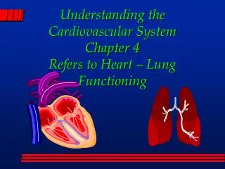
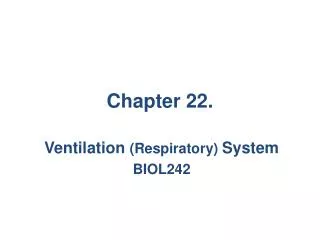
The structure & function of the respiratory system. What is gas exchange ?. The process of oxygen and carbon dioxide moving between the lungs and the blood. . What are the lungs?. Lungs are the organs of gas exchange. .

E N D
What is gas exchange? The process of oxygen and carbon dioxide moving between the lungs and the blood. What are the lungs? Lungs are the organs of gas exchange. The right lung is a little bigger than the left lung. This is because the left lung has to fit around the heart making it slightly smaller. The lungs are split into lobes The left lung is split into 2 lobes while the right lung is split into 3 lobes. Lungs have a very spongy texture and have a large surface area.
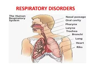
Trachea Ribs Alveoli Bronchi Bronchioles Lung Intercostal Muscles Diaphragm
Inhalation/ Inspiration (breathing in) Intercostal muscles contract Ribs move up and out Diaphragm contracts Diaphragm moves down / flattens (Thoracic) cavity increases in size Pressure surrounding lungs lowers compared with atmospheric pressure (a vacuum is created) Air rushes into lungs (down the trachea) AQA EXAM ANSWER!!! Inhalation is an ACTIVE process
Exhalation/ Expiration (breathing out) Intercostal muscles relax Ribs move down and in Diaphragm relaxes Diaphragm moves up (Thoracic) cavity decreases in size Pressure surrounding lungs increases compared with atmospheric pressure Air rushes out lungs (up the trachea) Exhalation is a PASSIVE process
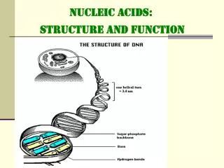
Lung structure • Gases are exchanged between the atmosphere and the blood through the respiratory surfaces of the lungs via diffusion. • The lungs consist of a mass of tiny air sacs called ‘alveoli’ supplied by many air tubes and blood vessels. • The alveoli are the site of gas exchange where O₂ and CO₂ diffuse across its thin walls. • They are just one cell thick and covered in capillaries • They increase surface area for maximum gas exchange
The Trachea Image taken from past exam paper! • A channel for air to flow to and from the bronchi • Glands produce a mucus lining which traps dust and bacteria • The beating of cilia on its surface move the mucus (and the dust/ bacteria trapped in it) to the pharynx for swallowing, preventing damage to lung tissue and dust, dirt, foreign bodies getting into lungs • The C-shaped rings of cartilage support the wall of the trachea keeping it permanently open to allow breathing • Bronchus - one goes to each lung - similar in structure to the trachea but narrower
Lung Capacity Definitions Tidal Volume The volume of air breathed in or out during one ventilation cycle Inspiratory Reserve Volume is the maximal volume of additional gas that can be inhaled through forcible inspiration following a normal inspiration.
Expiratory Reserve Volume is the additional volume of gas exhaled with maximum effort at the end of a normal, quiet expiration. Residual Volume is the volume of gas that remains in the lungs following a maximal expiration. Vital Capacity is the maximum volume of gas that can be forced out from the lungs at the end of maximum inhalation. Peak Expiratory Flow Rate is the measurement of the maximum rate of airflow attained during a vital capacity determination.
Measuring lung capacity – use a spirometer Inspiratory reserve volume Vital capacity Tidal volume during exercise Tidal volume at rest Expiratory reserve volume Residual volume
Normal lung capacity values • breathing rate 12–15 breaths per min • tidal volume 400–500 cm3 • vital capacity (male) 4.8 dm3 • vital capacity (female) 3.1 dm3 • peak flow 400–600 dm3 min−1 You MUST be able to recite these values for the exam!
Who has a larger vital capacity? Vs Why?
To get vital capacity To get tidal volume 1. Breathe in as far as you can through your nose then out as far as you can through your mouth. 1. Breathe normally into the spirometer 3 times. 2. Divide your reading by 3 to get an average tidal volume. 2. Repeat 2 more times and use the highest value. To use a spirometer What happens to each of the volumes with exercise?
Exercise & Vital Capacity • Regular exercise increases the vital capacity, here’s why: • Elasticity of chest wall increases • Chest wall able to expand (and recoil) more during exercise • Elasticity of lungs increases • Lungs able to expand (and recoil) more during exercise