Download
1 / 170
1.98k likes | 3.29k Vues
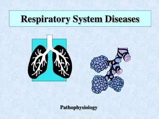
Diseases of the Respiratory System. Pathology Department of SiChuan University Su Xueying. Normal structure of the respiratory tract. The lower respiratory tract. Respiratory mucosa.

E N D
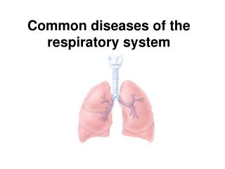
Diseases of the Respiratory System Pathology Department of SiChuan University Su Xueying
The lower respiratory tract
The respiratory system disease is very common • Emvironmental factors is important
Major aetiological factors in respiratory disease • Emvironmental Smoking Lung cancer Chronic bronchitis and emphysema Susceptibility to infection Air pollution Chronic bronchitis Susceptibility to infection Infection Influenza Pneumonia Tuberculosis Occupation Lung cancer Mesothelioma • Genetic Cystic fibrosis Some asthma
OUTLINE 1.Pulmonary Infections Bacteria Pneumonias Atypical Pneumonias Tuberculosis 2.Chronic Obstractive Lung Diseases (COPD) Emphysema Chronic Bronchitis 3.Bronchiectasis 4.Cor Pulmonale 5.Lung tumors
OUTLINE 1.Pulmonary Infections Bacteria Pneumonias Atypical Pneumonias Tuberculosis 2.Chronic Obstractive Lung Diseases (COPD) Emphysema Chronic Bronchitis 3.Bronchiectasis 4.Cor Pulmonale 5.Lung tumors
Definition Bacteriapneumonia is due to bacteria infection affecting distal airways, especially alveoli, with formation of an inflammatory exudate. often follows a viral upper respiratory tract infection
Streptococcus pneumoniae (pneumococcus) • Staphylococcus • Haemophilus influenzae • Klebsiella pneumoniae • Moraxella catarrhalis
Lobar pneumonia congestion stage red hepatization gray hepatization resolution Bronchopneumonia
Lobar pneumonia • Affects a large part, or the entirety of a lobe, frequently unilateral • Affects otherwise healthy adults between 20 and 50 years of age, males more than females • 90% due to Streptococcus pneumoniae
Stage of congestion Red, edematous
Red hepatization • Red • Solid • Consistency resembling fresh liver
Gray hepatization • Dry • Pale • Firm
Symptoms • High fever • Chills • Chest pain • Mucopurulent cough • with/without hemoptysis (rusty sputum) • Dyspnea
Bronchopneumonia(Lobular pneumonia) • Patchy consolidation • Centred on bronchioles or bronchi • Usually in infancy or old age • Usually secondary to pre-existing disease • Fever, cough
Outcomesof Pneumonia • Complete recovery • Complications developed Abscess formation Empyema Bacteremic dissemination • Organization
Diagnosis & Therapy Physical examination X-ray Blood culture Penicillin or other sensitive antibiotic treatment
Diagnosis & Therapy Physical examination X-ray Blood culture Penicillin or other sensitive antibiotic treatment
OUTLINE 1.Pulmonary Infections Bacteria Pneumonias Atypical Pneumonias Tuberculosis 2.Chronic Obstractive Lung Diseases (COPD) Emphysema Chronic Bronchitis 3.Bronchiectasis 4.Cor Pulmonale 5.Lung tumors
Atypical pneumonia • The concept was set forth in 1938 • The clinical course is unlike the “typical” bacteria pneumonia
Causes mycoplasma virus chlamydia
Gross morphology Red, congested Patchy or whole lobes
Microscopic characteristic the inflammatory reaction is largely confined within the walls of the alveoli, the septa are widened and edematous with mononuclear cells infiltration--- interstitial pneumonia