Download
1 / 63
630 likes | 655 Vues
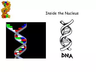
Delve into the fascinating world of DNA, its structure, and transformation through Griffith, Avery, and Hershey-Chase experiments. Understand the significance of nucleotides, base pairing, and the DNA double helix.

E N D
Interest Grabber Section 12-1 • Order! Order! • Genes are made of DNA, a large, complex molecule. DNA is composed of individual units called nucleotides. Three of these units form a code. The order, or sequence, of a code and the type of code determine the meaning of the message. 1. On a sheet of paper, write the word cats. List the letters or units that make up the word cats. 2. Try rearranging the units to form other words. Remember that eachnew word can have only three units. Write each word on your paper, and then add a definition for each word. 3. Did any of the codes you formed have the same meaning? 4. How do you think changing the order of the nucleotides in the DNA codon changes the codon’s message? Go to Section:
Section Outline Section 12-1 • 12–1 DNA A. Griffith and Transformation 1. Griffith’s Experiments 2. Transformation B. Avery and DNA C. The Hershey-Chase Experiment 1. Bacteriophages 2. Radioactive Markers D. The Structure of DNA 1. Chargaff’s Rules 2. X-Ray Evidence 3. The Double Helix Go to Section:
Objectives • Be able to define transformation, bacteriophage, nucleotide, and base pairing. • Be able to describe the Griffith, Avery, and Hershey-Chase experiments. • Be able to explain what scientists discovered about the relationship between genes and DNA. • Be able to explain the overall structure of the DNA molecule.
Vocabulary Words • Transformation – process in which one strain of bacteria is changed by a gene or genes from another strain of bacteria • Bacteriophage – a kind of virus that infects and kills bacteria • Nucleotide – monomer of nucleic acids made up of a 5-carbon sugar, a phosphate group, and a nitrogenous base • Base pairing – principle that bonds in DNA can form only between adenine and thymine and between guanine and cytosine
Griffith and Transformation • Griffith (mice injected with bacteria) – genetic information could be transformed from one bacterium to another
Figure 12–2 Griffith’s Experiment Section 12-1 Heat-killed, disease-causing bacteria (smooth colonies) Harmless bacteria (rough colonies) Harmless bacteria (rough colonies) Control(no growth) Heat-killed, disease-causing bacteria (smooth colonies) Disease-causing bacteria (smooth colonies) Dies of pneumonia Dies of pneumonia Lives Lives Live, disease-causingbacteria (smooth colonies) Go to Section:
Figure 12–2 Griffith’s Experiment Section 12-1 Heat-killed, disease-causing bacteria (smooth colonies) Harmless bacteria (rough colonies) Harmless bacteria (rough colonies) Control(no growth) Heat-killed, disease-causing bacteria (smooth colonies) Disease-causing bacteria (smooth colonies) Dies of pneumonia Dies of pneumonia Lives Lives Live, disease-causingbacteria (smooth colonies) Go to Section:
Avery and DNA • Avery (“juice” from heat-killed bacteria and enzymes) – DNA is the nucleic acid that stores and transmits the genetic information from one generation of an organism to the next
Alfred Hershey- Martha Chase • Hershey-Chase – genetic material of the bacteriophage is DNA, not protein
Figure 12–4 Hershey-Chase Experiment Section 12-1 Bacteriophage with phosphorus-32 in DNA Phage infectsbacterium Radioactivity inside bacterium Bacteriophage with sulfur-35 in protein coat Phage infectsbacterium No radioactivity inside bacterium Go to Section:
Figure 12–4 Hershey-Chase Experiment Section 12-1 Bacteriophage with phosphorus-32 in DNA Phage infectsbacterium Radioactivity inside bacterium Bacteriophage with sulfur-35 in protein coat Phage infectsbacterium No radioactivity inside bacterium Go to Section:
Figure 12–4 Hershey-Chase Experiment Section 12-1 Bacteriophage with phosphorus-32 in DNA Phage infectsbacterium Radioactivity inside bacterium Bacteriophage with sulfur-35 in protein coat Phage infectsbacterium No radioactivity inside bacterium Go to Section:
Figure 12–5 DNA Nucleotides Section 12-1 Purines Pyrimidines Adenine Guanine Cytosine Thymine Phosphate group Deoxyribose Go to Section:
Percentage of Bases in Four Organisms Section 12-1 Source of DNA A T G C Streptococcus 29.8 31.6 20.5 18.0 Yeast 31.3 32.9 18.7 17.1 Herring 27.8 27.5 22.2 22.6 Human 30.9 29.4 19.9 19.8 Go to Section:
Sugar-Phosphate Backbone and Chargaff’s Rule • Chargaff’s Rules: If there are a certain number of cytosines, there will be about the same number of guanines. Same with A’s and T’s.
Rosalind Franklin 1950 X-Ray Diffraction • Clues from the X-Ray • Coiled (forming Helix) • Double-stranded • Nitrogeneous bases are in the center
Watson & Crick • Francis Crick – British physicist • James Watson – American Biologist • Building a 3D model of DNA • Franklin’s X-Ray opened their eyes to the Double Helix • Watson and Crick’s model of DNA was a double helix, in which two strands were wound around each other.
Figure 12–7 Structure of DNA Section 12-1 Nucleotide Hydrogen bonds Sugar-phosphate backbone Key Adenine (A) Thymine (T) Cytosine (C) Guanine (G) Go to Section:
List the conclusions and explain how each of these scientists derived the conclusions: Griffith Avery Hershey and Chase Why did Hershey and Chase grow viruses in cultures that contained both radioactive phosphorus and radioactive sulfur? What might have happened if they only used one? How did Watson and Crick’s model explain why there are equal amounts of thymine and adenine in DNA? Questions
Interest Grabber Section 12-2 • A Perfect Copy • When a cell divides, each daughter cell receives a complete set of chromosomes. This means that each new cell has a complete set of the DNA code. Before a cell can divide, the DNA must be copied so that there are two sets ready to be distributed to the new cells. Go to Section:
Interest Grabber continued Section 12-2 1. On a sheet of paper, draw a curving or zig-zagging line that divides the paper into two halves. Vary the bends in the line as you draw it. Without tracing, copy the line on a second sheet of paper. 2. Hold the papers side by side, and compare the lines. Do they look the same? 3. Now, stack the papers, one on top of the other, and hold the papers up to the light. Are the lines the same? 4. How could you use the original paper to draw exact copies of the line without tracing it? 5. Why is it important that the copies of DNA that are given to new daughter cells be exact copies of the original? Go to Section:
Section Outline Section 12-2 • 12–2 Chromosomes and DNA Replication A. DNA and Chromosomes 1. DNA Length 2. Chromosome Structure B. DNA Replication 1. Duplicating DNA 2. How Replication Occurs Go to Section:
ProkaryoticChromosome Structure Section 12-2 • No Nucleus Chromosome E.coli bacterium Bases on the chromosome Go to Section:
Most have one circular chromosome located in the cytoplasm with some plasmids (circular DNA molecule found in bacteria) as well E.Coli (1.6μm diameter) 4,639,221 base pairs 1.6mm long Like packing 300m of rope in your backpack Prokaryote DNA
Eukaryotes and DNA • 1000 times more base pairs than bacterial DNA • Smallest human chromosome has 30 million base pairs of DNA • How do eukaryotes fit all that DNA in its nucleus?
Figure 12-10 Chromosome Structure of Eukaryotes Section 12-2 Nucleosome Chromosome DNA double helix Coils Supercoils Histones Go to Section:
DNA to Chromosomes • Vocabulary • Chromatin - granular material (uncondensed) within the nucleus; consists of DNA tightly coiled around proteins • Chromosomes – condensed chromatin • Histone - globular protein molecule around which DNA is tightly coiled in chromatin
DNA Replication • During DNA replication, the DNA molecule separates into two strands, then produces two new complementary strands following the rules of base pairing. Each strand of the double helix of DNA serves as a template, or model, for the new strand.
Enzymes unwind DNA • Enzymes split “unzip” double helix • The enzyme, DNA polymerase, finds and attaches the corresponding N-base • Each “old” stand serves as a template and is matched up with a new stand of DNA • New helixes wind back up.
Figure 12–11 DNA Replication Section 12-2 Original strand DNA polymerase New strand Growth DNA polymerase Growth Replication fork Replication fork Nitrogenous bases New strand Original strand Go to Section:
DNA Replication A – C – T – T – G – G – A – C T – G – A – A – C – C – T - G
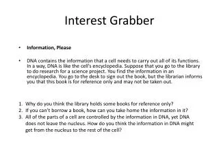
Interest Grabber Section 12-3 • Information, Please • DNA contains the information that a cell needs to carry out all of its functions. In a way, DNA is like the cell’s encyclopedia. Suppose that you go to the library to do research for a science project. You find the information in an encyclopedia. You go to the desk to sign out the book, but the librarian informs you that this book is for reference only and may not be taken out. 1. Why do you think the library holds some books for reference only? 2. If you can’t borrow a book, how can you take home the information in it? 3. All of the parts of a cell are controlled by the information in DNA, yet DNA does not leave the nucleus. How do you think the information in DNA might get from the nucleus to the rest of the cell? Go to Section:
Section Outline Section 12-3 • 12–3 RNA and Protein Synthesis A. The Structure of RNA B. Types of RNA C. Transcription D. RNA Editing E. The Genetic Code F. Translation G. The Roles of RNA and DNA H. Genes and Proteins Go to Section:
Messenger RNA Ribosomal RNA Transfer RNA Bringamino acids toribosome Combine with proteins tRNA mRNA Carry instructions rRNA DNA Ribosome Ribosomes Concept Map Section 12-3 RNA can be also called which functions to also called which functions to also called which functions to from to to make up Go to Section:
RNA and Protein Synthesis • Codon - three-nucleotide sequence on messenger RNA that codes for a single amino acid • Anticodon - group of three bases on a tRNA molecule that are complementary to an mRNA codon
Transcription Occurs in the nucleus Formation of a single strand of messenger RNA from DNA Translation Occurs on ribosomes Cell uses the information on mRNA to assemble amino acids in the proper order to form specific proteins Protein Synthesis: Two Parts
Transcription • Occurs in nucleus • Enzymes unwind DNA • Enzymes split “unzip” double helix • RNA polymerase binds to promoter sequence (signal) on DNA • RNA polymerase transcribes a single strand of mRNA
Figure 12–14 Transcription Section 12-3 Adenine (DNA and RNA) Cystosine (DNA and RNA) Guanine(DNA and RNA) Thymine (DNA only) Uracil (RNA only) RNApolymerase DNA RNA Go to Section:
Figure 12–17 The Genetic Code Section 12-3 Proteins are made by joining amino acids into long chains called polypeptides. Each polypeptide contains a combination of any or all of the 20 different amino acids. The genetic code shows the amino acid to which each of the 64 possible codons corresponds. There is one codon, AUG, that can either specify methionine, or serve as the initiation, or “start”, for protein synthesis. There are three “stop” codons that do not code for any amino acid.
Figure 12–18 Translation Section 12-3 Nucleus Messenger RNA Messenger RNA is transcribed in the nucleus. mRNA Lysine Phenylalanine tRNA Transfer RNA The mRNA then enters the cytoplasm and attaches to a ribosome. Translation begins at AUG, the start codon. Each transfer RNA has an anticodon whose bases are complementary to a codon on the mRNA strand. The ribosome positions the start codon to attract its anticodon, which is part of the tRNA that binds methionine. The ribosome also binds the next codon and its anticodon. Methionine Ribosome Start codon mRNA Go to Section:
Figure 12–18 Translation (continued) Section 12-3 The Polypeptide “Assembly Line” The ribosome joins the two amino acids—methionine and phenylalanine—and breaks the bond between methionine and its tRNA. The tRNA floats away, allowing the ribosome to bind to another tRNA. The ribosome moves along the mRNA, binding new tRNA molecules and amino acids. Growing polypeptide chain Ribosome tRNA Lysine tRNA mRNA Completing the Polypeptide The process continues until the ribosome reaches one of the three stop codons. The result is a growing polypeptide chain. mRNA Translation direction Ribosome Go to Section:
Questions • 1. What happens during DNA replication? • 2. List and describe the three main types of RNA. • Describe the interactions between DNA, RNA, and • proteins during each part of protein synthesis? • 4. Describe the main difference between RNA and • DNA.
Interest Grabber Section 12-4 • Determining the Sequence of a Gene • DNA contains the code of instructions for cells. Sometimes, an error occurs when the code is copied. Such errors are called mutations. Go to Section:
Interest Grabber continued Section 12-4 1. Copy the following information about Protein X: Methionine—Phenylalanine—Tryptophan—Asparagine—Isoleucine—STOP. 2. Use Figure 12–17 on page 303 in your textbook to determine one possible sequence of RNA to code for this information. Write this code below the description of Protein X. Below this, write the DNA code that would produce this RNA sequence. 3. Now, cause a mutation in the gene sequence that you just determined by deleting the fourth base in the DNA sequence. Write this new sequence. 4. Write the new RNA sequence that would be produced. Below that, write the amino acid sequence that would result from this mutation in your gene. Call this Protein Y. 5. Did this single deletion cause much change in your protein? Explain your answer. Go to Section:
Section Outline Section 12-4 • 12–4 Mutations A. Gene Mutations B. Chromosomal Mutations Go to Section: