Anatomy and Physiology 2 Hours
750 likes | 1.16k Vues
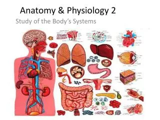
Anatomy and Physiology 2 Hours. Lynn E. Lawrence, CMSgt (ret), CPOT, ABOC. Tear Film Layers. oil. aqueous. snot. What functions does each layer of the tear perform?. A complex mixture of proteins, mucins, and electrolytes coated by a lipid layer. Healthy Tears. Antimicrobial proteins

Anatomy and Physiology 2 Hours
E N D
Presentation Transcript
Anatomy and Physiology 2 Hours Lynn E. Lawrence, CMSgt (ret), CPOT, ABOC
Tear Film Layers oil aqueous snot What functions does each layer of the tear perform?
A complex mixture of proteins, mucins, and electrolytes coated by a lipid layer Healthy Tears • Antimicrobial proteins • Growth factors & suppressors of inflammation • Soluble mucin helps stabilize tear film • Electrolytes for proper osmolarity (295-300) • pH slightly alkaline (7.4) Why is a tear salty tasting?
Left:Transillumination of eyelid showingmeibomian glands Lipid Secretion: Meibomian Glands Right: Secretion of lipid at lid margin • The lipid layer restricts evaporation to 5-10% of tear flow • Also helps lubricate Where does a contact lens rest?
Transillumination ofmeibomian glands (WC Posey, Diseases of the Eye, 1902) Lipid Secretion: Meibomian Glands What eye is this? How does the lipid layer aid in contact lens wear? (Transillumination image from Dry Eye and Ocular Surface Disorders, 2004)
Anatomy What function does the pupil have?
The Eyelid • 7 Layers of the eyelids 1. Skin-thinnest layer 2. Subcutaneous connective tissue 3. Striated Muscle 4. Sub-muscular connective tissue 5. Tarsal plate or fibrous layer 6. Smooth muscle 7. Conjunctiva (Bulbar/Palpebral) How are Hordoleum and Chalazions treated?
Eyebrows and Eyelashes • Eyebrows • Thickened ridge of skin with short hairs • Diverts perspiration • Eyelashes • Also protects • Sebaceous glands at base of each lash are called Glands of Zeis which produce a lubricating fluid • Fluid can harden and clog the gland, producing a stye or painless chalazion. If painful and infected it is called an external hordeolum Cataracts (myotonia) are caused by?
Lacrimal Apparatus • Sometimes a person cannot produce natural tears that they might need some punctal plugs. What are the three main parts of the crystalline lens?
Anatomy and Physiology of the Eyeball 3 Layers • Fibrous Layer *Cornea *Sclera • Vascular Layer *Choroid *Ciliary body *Iris • Nerve Layer *Retina *Macula *Optic nerve What is the main function of each layer?
Sclera • White in color • Primary function is protection • Pierced posteriorly by the optic nerve • Acts as insertion points for the six EOMs • Junction between the cornea and sclera is called the Limbus What is the total power of the eye?
Conjunctiva • An epithelial membrane which covers the anterior sclera and continues to the back surfaces of the lids to form a conjunctival sac • Has blood vessels which can burst and cause subconjunctival hemorrhage • Three parts • Bulbar • Palpebral • Fornix - where bulbar and palpebral meet Name the mucus producing cell?
Cornea • Index of refraction is 1.37 • Approximately .5mm in thickness • Transparent Organ (no blood vessels / avascular) • Primary function is refraction of light rays • Refractive power approx + 45.00 D What is it called when blood vessels grow onto the cornea? What happens when a patient gets a scar in the visual pathway? What is the crossover point for the nasal optic nerves?
Cornea • Composed of 5 layers • Epithelium…24 hr healing • Outermost layer • 5 cell layers thick • Heals very quickly • Does not scar • Bowman's membrane- layer just under the epithelium NOTE: will scar • Stroma – middle tissue that forms 90% of the cornea • Descemet's membrane- thin elastic layer deep in the cornea • Endothelium - only one cell layer thick; lines undersurface of the cornea, where it regulates corneal water content What cranial nerve is tied to corneal sensations?
Endothelial detail with nuclei Confocal Scanner Epithelial detail with nuclei Which cells do not regenerate Epithelium or Endothelium?
Aqueous Humor • Manufactured by ciliary body • Characteristics: • Clear • Watery consistency (99% H2O) • Functions • Refraction of light • Intraocular Pressure (IOP) • Probably nourishes posterior surface of the cornea and the crystalline lens • Flows from posterior chamber through the pupil into the anterior chamber How does aqueous flow out of anterior chamber?
Crystalline Lens...approx 12-14 diopters of power • Functions • Refraction of light • Accommodation • Focus adjustment of the eye • Presbyopia is the loss in accommodation • First noticed around age 40. Due to a loss in flexibility of the lens Name the three main parts of the lens?
Crystalline Lens • 3 things happen during accommodation: • Pupils constrict • Eyes converge • Lens gets thicker • The crystalline lens contains a high degree of protein • Changes in the lens protein causes the lens to lose its transparency which is a condition termed "cataract" • Aphakia is the absence of a lens. It can be removed during cataract extraction How much focusing power does the lens have?
Iris • Most anterior portion of the vascular layer • Gives the eye its color, i.e. blue eyes, brown eyes, etc. • Consists of blood vessels, pigment and muscle tissue • Regulates light • Smaller with age What does the sphincter muscle control?
Ciliary Body • Located near the base of the iris and posterior to it • Composed of blood vessels and muscle fibers (ciliary muscle) • Cilliary process produces aqueous Ciliary body is attached to suspensatory ligaments called?
Vitreous Chamber • Functions: • Refraction of light • Internal support • Spots in vision may be floaters in the vitreous Post vitreous detachment How many chambers are inside the eye?
Nerve Layer - Retina • Visual Receptors are Cones and Rods • Cones • Produce color vision • Give improved acuity • Used in day vision = “Photopic” = normal and high levels of illumination • Rods…120 million • Produce black and white vision • Function in dim light = “Scotopic” = low level of illumination • Cones and Rods… 6 million • Used under mesopic vision = between scotopic and photopic • Both rods and cones are used. The _____ is the strongest refractive media and has about ____ diopters of power.
The retina (Cranial Nerve II) Pigment epithelium Vitreous The levator palpebrae raises the eyelid and is innervated by CN #?
Retina – 10 layers Outside of eye • Pigment epithelium • Rods • Cones • Outer plexiform layer • Horizontal cells • Bipolar cells • Amacrine cells • Inner plexiform layer • Ganglion cells • Nerve fiber layer Vitreous (inside of eye)
Identification of Retinal Layers NFL ILM GCL IPL OPL Stratus OCT™ IS/OS RPE/CC Choroid IPL: Inner Plexiform Layer OPL: Outer Plexiform IS/OS: Junction of inner and outer photoreceptor segments RPE: Retinal Pigment Epithelium CC: Choriocapillaris NFL: Nerve Fiber Layer ILM: Inner Limiting Membrane GCL: Ganglion Cell Layer Cross-sectional image of live tissue; a virtual biopsy
Nerve Layer - Retina • Optic nerve head (optic disc) • No receptors - physiological blind spot • Point of exit of optic nerve • Appears yellow compared to the orange retina What is Pars Plana?
Nerve Layer - Retina • Ora Serrata • Land mark attachment site for choroid and retina • Most anterior portion of retina • Nearly all rods How many layers are in the retina?
Anatomy and Physiology of the extraocular muscles • The Extra-ocular Muscles (EOM) • Organized into an umbrella-like bundle among the orbital fat, orbital blood vessels and nerves • Six muscles associated with eye movements • Superior rectus (S.R.) • Inferior rectus (I.R.) • Medial rectus (M.R.) • Lateral rectus (L.R.) • Superior oblique (S.O.) • Inferior oblique (I.O.) How many cranial nerves control these 6 muscles?
Extra Ocular Muscles What is the name of the point where the muscles come together?
Extraocular Muscles • Medial Rectus - Most powerful, adduction, CN III • Inferior Rectus - Primary is depression, CN III • Lateral Rectus - Abduction, CN VI • Superior Rectus - Primary is elevation Which muscle close the eye lid and is innervated by cranial #7?
Muscles and Function • LR6…SO4…3 • Rectus • Obliques • Intorsion • Extorsion • Elevation • Depression An obvious upward/superior deviation of the eye is called?
Extraocular Muscles • Superior Oblique (SO)-has 3 functions; intorsion, depression and abduction; innervated by the 4th (trochlear) cranial nerve • Inferior Oblique (IO)-3 functions; extorsion, elevation, and abduction; innervated by the 3rd (oculomotor) cranial nerve Proper alignment and muscle balance of the eyes is called?
Extraocular Muscles • Medial Rectus (MR)-moves the eye inward from the straight-ahead position (adduction); innervated by the 3rd (Oculomotor) cranial nerve • Lateral Rectus (LR)-moves the outward (abduction) from the straight-ahead position; innervated by the 6th (Abducens) cranial nerve • Inferior Rectus (IR)-3 functions; depression, extorsion, and adduction; innervated by the 3rd (Oculomoter) cranial nerve A definite and obvious turning of the eye is called?
Ocular Motility • Version - a conjugate movement of the 2 eyes. Both eyes remain parallel during the movement • Vergence - A disjunctive movement of the 2 eyes • Convergence • Near triad of accommodation, pupil constriction, and convergence • Divergence A constant tendency for the eyes to turn from the norm is called?
Muscle Balance Testing • Cover Test • Cover/uncover • Alternating cover • Hirschberg Test • Location of corneal reflex Which test checks for direction when using the cover test?
Extraocular Muscles • Medial Rectus • Toward the nose (adduction) • Lateral Rectus • Away from the nose (abduction) • Superior Rectus • Up;towards the nose (elevation) • Inferior Rectus • Down;away from the nose (depression) How many extra ocular muscles are there?
Extraocular Muscles • Superior Oblique • Rotates the top of the eye toward the nose;moves eye down • Inferior Oblique • Rotates the top of the eye away from the nose; moves eye up Where is the insertion points for these muscles?
Bony Orbit • Openings of the orbit • Purpose of openings • Transmit arteries and/or veins to and from the orbit • Transmit nerves to and from the orbit • Types of openings • Fissures (crevices/cracks) • Foramina (holes) • Major openings • Optic foramen - II cranial nerve - Optic Nerve • Supraorbital fissure - IV cranial nerve - Trochlear Nerve The transition zone between the sclera and the cornea is called?
Orbit • Frontal bone…forehead • Ethmoid bone…weakest • Palatine bone…smallest • Zygomatic bone…strongest • Lacrimal bone • Maxillary bone The conjunctiva has two divisions, they are?
Cranial Nerves LR6SO43 Muscles • Lateral rectus muscles #6 …abducens nerve • Superior Oblique #4 …trochlear nerve • All other muscles are controlled by #3 … oculomotor nerve Name the 3 chambers of the internal eye?
Anatomy Physiology • The Orbit - Bones, etc. • The Sinuses - Locations • Human Body Planes • External Structures - Eyelids - Conjunctiva - Eyelashes and Eyebrows • Lacrimal System The outer layer of the eyeball is called?
Visual Pathway Objectives • Define the visual pathway • Identify structures in the visual pathway • Testing used for the visual pathway • Identify defects within the visual pathway Anisometropia occurs when there is a _____________?
Visual Pathway • Visual pathway is the path taken by nerve impulses between the eye and the brain when the retina is stimulated by light Iseikonic Lenses are designed are designed for what?
Visual Pathway • Physical • Physiological • Psychological What causes your physiological blind spot?
Visual Pathway • Visual pathway has seven structures • Retina • Optic Nerve • Optic Chiasm • Optic Tract • Lateral Geniculate Body (LGB) • Optic Radiations • Visual Cortex …where vision occurs Aniseikonia occurs when an object viewed by one eye is _________?
Visual Pathway Antimetropia occurs when __________ ?
Retina • Divided into four quadrants like the brain • Fovea at exact center • Optic nerve head is located in nasal half • Each quadrant sees the exact opposite visual field What is an exudate? What is papilledema?
Optic Chiasm • Temporal fibers do not cross • Nasal fibers do cross • Some fibers from the macula cross while others do not What part of the brain does the vision occur?
Visual Field Defects Homonymous heminopia • Common types of field defects • Blind spots - Areas of blindness in the visual field • Hemianopsia - Blindness in one half of the visual field of one or both eyes • Homonymous - Involving the nasal half of the visual field of one eye and the temporal half of the visual field of the other eye • Incongruous - Incongruous homonymous Binasal defect An area of blindness within a visual field is called?