Download
1 / 63
640 likes | 782 Vues
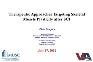
Therapeutic Approaches Targeting Skeletal Muscle Plasticity after SCI. Chris Gregory Assistant Professor Department of Health Sciences & Research Medical University of South Carolina Health Science Specialist Research Service Ralph H. Johnson VAMC July 17, 2012.

E N D
Therapeutic Approaches Targeting Skeletal Muscle Plasticity after SCI Chris Gregory Assistant Professor Department of Health Sciences & Research Medical University of South Carolina Health Science Specialist Research Service Ralph H. Johnson VAMC July 17, 2012
Disclosure of PI-RRTC Grant • James S. Krause, PhD, Holly Wise, PhD; PT, and Emily Johnson, MHA have disclosed a research grant with the National Institute of Disability and Rehabilitation Research. • The contents of this presentation were developed with support from an educational grant from the Department of Education, NIDRR grant number H133B090005. However, those contents do not necessarily represent the policy of the Department of Education, and you should not assume endorsement by the Federal Government.
Accreditation • The Medical University of South Carolina is accredited by the Accreditation Council for Continuing Medical Education (ACCME) to provide continuing medical education for physicians. The Medical University of South Carolina designates this live activity for a maximum of 1.0 AMA PRA Category 1 Credit(s)™. Physicians should claim only the credit commensurate with the extent of their participation in the activity. • In accordance with the ACCME Essentials &Standards, anyone involved in planning or presenting this educational activity will be required to disclose any relevant financial relationships with commercial interests in the healthcare industry. This information is listed below. Speakers who incorporate information about off-label or investigational use of drugs or devices will be asked to disclose that information at the beginning of their presentation. • The Center for Professional Development is an approved provider of the continuing nursing education by the South Carolina Nurses Association an accredited approver by the American Nurses Credentialing Center’s Commission on Accreditation
Disclosure of Presenter • Chris Gregory, PhD, PT, has disclosed research grants with the Department of Veteran’s Affairs, American Heart Association, South Carolina Spinal Cord Injury Research Fund and NIH/NINDS.
Prevent 2°Damage Axon Regeneration Create Bridges Replace Dead Cells
Outline • Skeletal muscle adaptations following SCI • Acute vs. Chronic • Secondary Health Conditions associated with alterations in skeletal muscle after SCI • Effects of exercise training on skeletal muscle after SCI • Health-related benefits of exercise in persons with SCI
Spinal Cord Injury Physical Inactivity / Muscle Unloading Skeletal Muscle ↓CV Function ↓Bone Density • Secondary Conditions: • NIDDM • CVD • Obesity • Dyslipidemia • Decreased fitness • Osteoporosis
Secondary conditions following SCI • Increased risk of cardiovascular disease(Kocina, 1997) • Leading cause of death after 20 yr DOI • ~90% > risk of heart attack relative to un-injured • ~228% > mortality risk from CVD • 46% of patients over 30 die from CVD • Decreased HDL • Increased risk of NIDDM (Bauman, 2000) • ~4x incidence of NIDDM after SCI • ~65% have impaired CHO metabolism vs 15% in un-injured population • Decreased bone density • ~50% reduced in 100% of population
Complete injury • Absence of sensory and motor function in the lowest sacral segment • Incomplete injury • Partial preservation of sensory and/or motor function below the neurologic level
ASIA Impairment Scale: • A No motor or sensory function in the lowest sacral segment • B Sensory but not motor function is preserved in the lowest sacral segment • C Less than ½ of the key muscles below the (single) neurological level have a grade 3 or better • D At least ½ of the key muscles below the (single) neurological level have a grade 3 or better 5 – Normal strength 4 – Full ROM with < normal strength against resistance 3 – Full ROM against gravity 2 – Movement with gravity eliminated 1 – Palpable contraction but no movement 0 – Total paralysis
Skeletal muscle adaptations following “Chronic” SCI Hillegass and Dudley 1999 Severe Muscle Atrophy (60-75%) Able-bodied male C5 complete SCI male
Quadriceps muscle CSA and total quadriceps muscle volume in SCI and able-bodied (AB) individuals. Olive J L et al. J Appl Physiol 2003;94:701-708
Skeletal muscle adaptations following “Chronic” SCI Conversion to predominately fast fibers Control SCI Martin et al. 1992 Gregory et al. 2003
Muscle Fatigue ~36% ~66% Bickel et al 2004
Muscle Fatigue Olive J L et al. J Appl Physiol 2003;94:701-708
Muscle Fatigue PCr recovery curves following NMES for (A) control and (B) SCI individual.
Muscle Injury • 25 ± 6 % • 2 ± 1 % Bickel et al 2004
Long-term SCI Specific Tension - force per unit area. Torque (Nm) = 3.74 x stimulated CSA (cm2) - 14.37, (R2 = 0.93, p < 0.05) Bickel et al. 2004
“Chronic” SCI Muscle • Severe muscle atrophy • Conversion to predominately fast fibers. • Increased fatigability • Increased susceptibility to injury • Force generating capacity (relatively) maintained
Skeletal muscle adaptations following “Chronic” SCI • Individuals years after SCI have been reported to have muscles that are small, highly fatigable and susceptible to damage; but still produce adequate force relative to their size. (Grimby 1976, Rochester et al. 1995, Hillegass and Dudley 1999 ) C5 complete SCI male Able-bodied male
Skeletal muscle adaptations following “Acute” SCI Pre-injury 12 weeks post-injury “Classic” Brown-Sequard syndrome
Muscle Atrophy MRI - 6, 12, 24 weeks after injury Thigh Muscles - Hamstring (-14%) - Adductors (-16%) - Quadriceps (-16%) Castro et. al. 1999a
Muscle Atrophy MRI - 6, 12, 24 weeks after injury Shank Muscles - TA (5% decrease, NS) - Soleus (-12%) - Gastrocnemius (-24%) Castro et. al. 1999a
Muscle Atrophy SCI vs. Controls
Muscle Atrophy Dudley et al. 1999
Fiber type * Data from m. Vastus Lateralis Castro et. al. 1999b
Enzyme changes GPDH SDH SDH - Kreb’s cycle (aerobic) GPDH - glycolytic enzyme (anaerobic) CSA Castro et. al. 1999b
Muscle Fatigue Increased muscle fatigue after SCI despite the alterations in muscle Castro et al 1999b Bickel et al 2004
“Acute” SCI Muscle • Muscle specific atrophy • Change in fiber type composition. • (IIa to IIx) • No decrease in % slow muscle in first 6 months • Energy supply/energy demand apparently unaltered. • Fatigability is increased compared to controls
Muscle AtrophyIncomplete SCI Relative differences in individual muscle CSA between the more-affected limb following incomplete SCI vs. control subjects.
“Chronic” SCI Bone SCI results in a dramatic loss of bone and a marked increase in fracture incidence • Chronic SCI results in a 50-70% demineralization and is correlated with duration of injury • Following complete SCI, bone loss proceeds at a rate of ~1% per week for the first 6-12 months • Microgravity = 0.25% /week • Bed rest = 0.1% /week • Menopause 3-5% /year Dauty et al. 2000; Modelesky et al. 2004
“Chronic” SCI Bone SCI results in a dramatic loss of bone and a marked increase in fracture incidence Modelesky et al. 2004
“Chronic” SCI Bone SCI results in a dramatic loss of bone and a marked increase in fracture incidence Distal Femur Proximal Tibia SCI Control Modelesky et al. 2004
“Chronic” SCI Bone Relationship between muscle volume and cortical bone volume and BMC remains strong SCI: r = 0.90 and 0.83 Modelesky et al. 2004
Vascular Response to SCI Femoral artery diameter Peak blood flow
So What? In the next several decades, a cure for SCI is a realistic possibility. Without a means to preserve the musculoskeletal integrity of paralyzed lower limbs, people injured today with SCI will be "inappropriate candidates" for reintroduction to standing and walking, should a cure be found.
Is it possible to elicit changes in muscle and bone years after SCI? • Early studies suggest an inability to increase muscle size in subjects years after injury • Other studies do report modest muscle hypertrophy (0-15%) following NMES cycling • Is this enough? • Limitations? • Program design
Resistance exercise increases muscle size & strength Aerobic/endurance exercise does not
SCI - Resistance Training 2 days/week x 6 months Electrical stimulation (30 Hz trains of 450 µsec biphasic pulses) 4 sets of 10 knee extensions Resistance - cuff weights All training performed at subjects’ homes
4 sets of 10 2 days per week for 8 weeks Total contractions = 640 Dudley et al. 1999
Molecular signals Effects of (NMES)-induced exercise on the expression and/or accumulation of mRNA for components of the muscle insulin-like growth factor (IGF)-I system Bickel et al 2003
SCI - Resistance Training “Chronic” Before 6 Months 3 Months After 6 months of training: Subjects were using between 14 - 30 pounds for resistance at the ankle. QF muscle size increased ~60% in both thighs.
SCI - Resistance Training “Chronic” Average AB muscle size (Castro et al. 1999) 62 58 52 49 37 34 • n = 5 • *p < 0.05, 3 Mo.Vs. Before • ‡ p<0.05, 6 Mo.vs.3 Mo. LEFT THIGH RIGHT THIGH Mahoney et al 2005
SCI - Resistance Training “Chronic”
SCI - Resistance Training “Chronic” FIGURE 1 . Axial MR Images of thigh (A) and knee extensors skeletal muscles (B) and %IMF (C) before and after RT + diet versus diet groups. Gorgey et al., Med & Sci Sports & Exer. 44(1):165-174
SCI - Resistance Training “Chronic” Electrically stimulated RT significantly lowered plasma glucose response during OGTT (n=5)
SCI - Resistance Training “Chronic”