Electron probe microanalysis - Scanning Electron Microscopy EPMA - SEM
UW- Madison Geoscience 777. Electron probe microanalysis - Scanning Electron Microscopy EPMA - SEM. Preface: What ’ s EPMA and SEM all about? How does Geology 777 work? What can you learn?. Updated 1/16/13. UW- Madison Geology 777.

Electron probe microanalysis - Scanning Electron Microscopy EPMA - SEM
E N D
Presentation Transcript
UW- Madison Geoscience 777 Electron probe microanalysis - Scanning Electron Microscopy EPMA - SEM Preface: What’s EPMA and SEM all about? How does Geology 777 work? What can you learn? Updated 1/16/13
UW- Madison Geology 777 You are taking this class because you need to be able to intelligently use one or both of the Department’s electron beam instruments -- the Cameca SX51 electron microprobe in Weeks 306 or the Hitachi S3400 SEM in Weeks 308. Both instruments work on the same physical principles, but have differences. Traditionally this class has focused primarily upon the electron microprobe, but over the past few years more material has been added to address specific issues with increased usage of the SEM. It is a work in progress… Why?
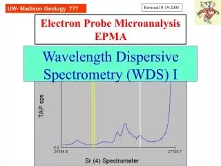
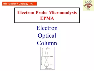
UW- Madison Geology 777 EPMA is a tool to get precise and accurate quantitative chemical analyses of micron-size domains of our samples. A focused beam (“spot”) of high energy electrons interact with the atoms in the sample, yielding X-rays (and other signals), which we quantify and compare with counts from standards. It is nominally non-destructive. EPMA - what is it?
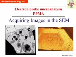
UW- Madison Geology 777 SEM is a tool to produce images -- pictures -- of our samples. A rastered (scanned) beam of high energy electrons sweeps across the surface, interacting with the atoms in the sample, yielding backscattered electrons, secondary electrons, auger electrons, and in some cases photons in the visible light range (CL). It is nominally non-destructive. SEM - what is it?
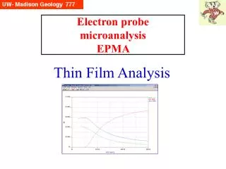
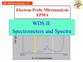
UW- Madison Geology 777 This technique has its own characteristics, strengths, weaknesses. It pays to consider whether it is the best technique to get the information you need. It is a micro-technique, and for multiphase samples provides discrete compositions, not the bulk composition. Under “normal operataing conditions”, it samples volumes (widths-depths) on the order of ~1-3 um, limiting its usefulness for smaller inclusions or thin films. It provides major and minor element quantification, and has limited capacity for trace element analysis. (What do you mean by “trace”?) Despite being non-destructive, samples need to be mounted and polished; they can be reanalyzed many times. It is relatively inexpensive and accessible Some degree of complexity; there can be a sharp learning curve EPMA - is it for me?
UW- Madison Geology 777 This technique is rather simple and one can learn the essentials in a short time. It provides images easily, though one needs to understand the various parameters (e.g. working distance, resolution, etc) to not make mistakes compromising image quality. Samples may be imaged with little or no preparation (coating, mounting+polishing), though this may complicate detailed examination. It is very easy to make mistakes using the easy EDS software, especially for attempts to get chemistry of small particles. SEM - is it for me?
UW- Madison Geology 777 The goal is to provide useful background information to make SEM and EPMA less a ‘black box’ for you and to help you make better decisions about how to analyze your samples, and to understand when data is good and when it is not. This class will provide the basic instructions for the use of our Hitachi SEM and Thermo-Fisher EDS. It will point out errors that can occur with EDS spectral interpretation. This course provides some directed exercises with our Cameca SX51. The electron probe is much more complicated than the SEM and experience has shown that individualized training is the best way to go. Which means this happens only when the student has his/her samples ready to analyze do we set up a 4-8 hour appointment. Goal of this course
UW- Madison Geology 777 Weekly class meetings: ~1.5 hours, discussion of assigned materials (PPT presentations, readings); students will be responsible for many of the readings Weekly quiz: at start of each class, on the assigned material Weekly labs: ~2 hours. Complete lab report and turn in following week Weekly assignments: Calculations and computer exercises. Each student will present an assigned published paper How this course is structured
UW- Madison Geology 777 Use for Reference--In Library on Reserve Goldstein et al, 3rd Edition. 2003 New:$75
UW- Madison Geology 777 Also On Reserve in Geo Library Reed (1996) 201 pages Paper: New:$36 Hard:New: $95 Used $80 Reed (1993) Paper: New:$55 Used:$35? Hard:New ~$95
UW- Madison Geology 777 Simple assumptions: We have stuck our sample in epoxy, cut and polished it (or made a thin section epoxied to a glass slide and polished it). There are standards, either user-supplied, or in the probe lab. We sign up on the schedule, get some “on the job” training, and analyze our samples. We return to our office with the data we need ready to show our advisor. EPMA - “ideal” case
UW- Madison Geology 777 Optimal case for “easy” EPMA: The samples are flat, well polished, conductive, non-porous, infinitely thick (to e- beam), homogeneous, clean. Standards exist and have the same 5 features. Materials are oriented at 90° to the electron beam (not tilted). Background positions well chosen with no peak or background interferences (in both unknown and standard). Detector pulse distribution well centered. Constants for matrix correction (e.g. mass absorption coefficients) well known. Sample is not able to be altered by beam. The devil in the details
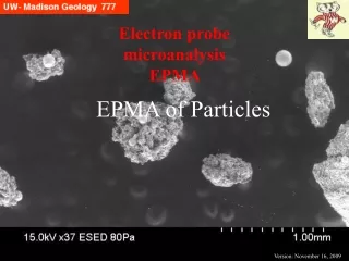
UW- Madison Geology 777 Possible complications: The actual materials being probed are scratched or etched, insulators, porous, multiphase (eutectic) assemblages, with polishing oil in pores. 1” round has surface that is not normal to walls, resulting in tilted surface to electron beam. Background and peak positions have interferences. Detector pulse distribution on standard depressed (cut off) on low end. Mass absorption coefficients poorly known. Specimen is a particle -- or a thin film. Specimen is hydrous or sensitive to alteration or damage by the beam electrons. A main goal of this course is for you to understand when the optimal conditions are met -- and when they are not -- and if there is a way to make this thing work! EPMA – real case details
UW- Madison Geology 777 How to trust the results?: Evaluate “secondary” standards. “Should” get 100 wt% totals (98.5-101). Evaluate stoichometry if able to. How to get the best results: Get the sample preparation right. Have multiple standards for difficult samples. Take some time at the start: Don’t be in a hurry with a new sample/suite of elements. Do wavescans first to consider peak and background interferences. EPMA - so what to do?