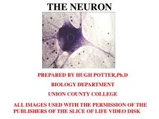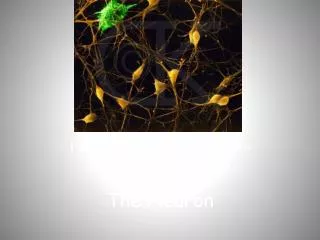
Information Flow and the Neuron
440 likes | 579 Vues
Information Flow and the Neuron. Chapter 37. Learning Objectives. List the 4 functions of neural cells Describe the two types of neuronal cells Diagram the basic nerve cell and myelination Explain a basic neuronal circuit (SAINE). Learning Objectives.

Information Flow and the Neuron
E N D
Presentation Transcript
Information Flow and the Neuron Chapter 37
Learning Objectives • List the 4 functions of neural cells • Describe the two types of neuronal cells • Diagram the basic nerve cell and myelination • Explain a basic neuronal circuit (SAINE)
Learning Objectives • Describe the flow of an action potential across the axon of a neuron, including the action of Na+ and K+ gated ion channels, and understand how myelination adds to the transmission of the action potential • Diagram the action of neurotransmission at synapses • List the types of medications that can affect neurotransmission in humans
37.1 Neurons and the Nervous System • Neurons are cells specialized for the reception and transmission of informational signals • Neurons are supported structurally and functionally by glial cells • Neurons communicate via synapses
4 Functions of the Animal Nervous System 1. Reception • Receives information about conditions in internal and external environment 2. Transmission • Transmits message along neurons
4 Functions of the Animal Nervous System 3. Integration • Integrates information to formulate appropriate response 4. Response • Sends out signals to muscles or glands
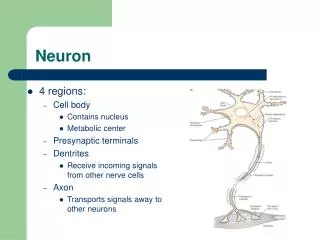
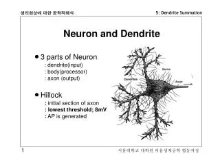
Neuron Structure • Dendrites • Receive information • Conduct signals toward the cell body • Axons • Conduct signals away from the cell body to another neuron or an effector
Dendrites Cell body Axon Nucleus Axon terminals Axon hillock Fig. 37.3, p. 849
SAINE • Sensory • Afferent • Inter Neuron • Efferent
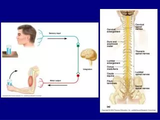
A Basic Neuron Circuit An afferent neuron, an interneuron, and an efferent neuron make up a basic circuit • Circuits combine into networks that interconnect the peripheral and central nervous systems • The Sensory, afferent, and efferent are the PNS • The Interneurons are the CNS
External, for example, light, movement; or internal, for example, change in blood pressure, change in body temperature Stimulus Sensory receptors of afferent neurons in external or internal organs detect stimulus. Reception Peripheral nervous system (PNS) Afferent (sensory) neurons Message travels along neuron. Transmission Central nervous system (CNS) Interneurons Neural messages sorted and interpreted. Integration Interneurons Message travels along neuron. Transmission Efferent neurons Neural messages from efferent neurons transmitted to effectors. Response Effectors Action Fig. 37.2, p. 848
Myelin sheath of Schwann cell Node of Ranvier Myelin sheath of Schwann cell Cytoplasm of axon Axon of neuron Plasma membrane of axon Fig. 37.5, p. 850
How do neurons “work” • Step 1- Resting potential • Step 2- Threshold potential • Step 3- Action potential • Step 4- Refractory period (hyperpolarization)
Membrane Potential of a Cell • Unequal distribution of positive and negative charges on either side of the membrane • Establishes a potential difference across the membrane
37.2 Signal Conduction by Neurons • Resting potential is the unchanging membrane potential of an unstimulated neuron – it is approx -70 mVolts • Membrane potential changes from negative to positive during an action potential • Action potential is produced by ion movements through the plasma membrane (Na+ and K+)
Resting Potential of Neurons (1) Na+/K+active transport pump • Sets up concentration gradients of Na+ ions (higher outside) and K+ ions (higher inside) (2) Open channel (passive) allows K+ to flow out freely (3) Negatively charged molecules (proteins) inside cell can’t pass through membrane (semipermeable)
Voltage-gated K+ channel (closed) Voltage-gated Na+ channel (closed) Axon plasma membrane Na+/K+ pump 3 Na+ out Na+ K+ A– K+ A– Na+ A– 2 K+ in Axon A– A– A– A– A– A– A– Anions (negatively charged proteins, nucleic acids, and other large molecules) that cannot pass through membrane Charged Particle Concentrations (mM) Inside Outside Na+ 150 15 K+ 5 150 0 100 A– Fig. 37-8, p. 853
Action Potential • Generated when stimulus pushes resting potential to threshold value • Voltage-gated Na+and K+ channels open in the plasma membrane • Inward flow of Na+ changes membrane potential from negative to positive peak • Potential falls to resting value as gated K+ channels allow ion to flow out
Refractory period Peak of action potential Depolarization Repolarization Membrane potential (mV) Threshold potential Resting potential Stimulus Hyperpolarization Time (msec) Fig. 37.9, p. 853
Outside cell Na+ channel K+ channel Activation gate Na+ Activation gate Na+ K+ Inactivation gate Cytoplasm Membrane potential (mV) Threshold potential Resting potential Time (msec) 1. A stimulus raises the membrane potential to threshold. The activation gate of the Na+ channel opens. Fig. 37-10a (1), p. 854
Na+ Na+ K+ Membrane potential (mV) Time (msec) 2. Above the threshold, more Na+ channels open and Na+flows inward along its concentration gradient, raising the membrane potential toward the peak of the action potential. Fig. 37-10a (2), p. 854
K+ Na+ Na+ Membrane potential (mV) Time (msec) 3. As the action potential reaches its peak, the inactivation gate of the Na+ channel closes and the K+ channel activation gate opens, allowing K+ ions to flow outward. Fig. 37-10a (3), p. 854
Membrane potential (mV) Time (msec) 4 The outward flow of K+ along its concentration gradient causes the membrane potential to begin to fall. Fig. 37.10b (1), p. 855
Membrane potential (mV) Time (msec) 6 Closure of the K+ activation gate stabilizes the membrane potential at the resting value. Fig. 37.10b (3), p. 855
Summary • Rest- active pump Na+ out, passive pump K+ in; more positive outside • Stimulation causes active gated channel to open and Na+ goes in until… • Threshold- all Na+ open quickly rises to peak • Peak- Na+ gated close, K+ gated open to let K+ out • Hyperpolarization- gates close and the pumps take over to reach resting potential again.
Propagation of Action Potential • Action potentials are initiated by dendrite, and build in the axon hillock. • Action potentials move along an axon as the ion flows generated in one segment depolarize the potential in the next segment
Adjacent area that was brought to threshold by local current flow; now active at peak of action potential Previous active area returning to resting potential; no longer active because of refractory period Time = 1 New adjacent inactive area into which depolarization is spreading; will soon reach threshold Remainder of axon still at resting potential – – – – – – Membrane potential (mV) Fig. 37.11b, p. 856
Refractory Period • Action potentials are prevented from reversing direction by a brief refractory period • A segment of membrane that has just generated an action potential can’t produce another for a few milliseconds
Saltatory Conduction • In myelinatedaxons, ions can flow across the plasma membrane only at nodes where the myelin sheath is interrupted • Action potentials skip rapidly from node to node
Synapses • Site where a neuron communicates with another neuron or effector • Presynaptic cell • Neuron that sends a signal • Postsynaptic cell • Neuron that receives a signal
2 Types of Synapses • Electrical synapse • Impulses pass directly from sending cell to receiving cell • Chemical synapse • Neurotransmitter released by presynaptic cell diffuses across synaptic cleft • Binds to receptors in the plasma membrane of postsynaptic cell
Neurotransmitters • Released into synaptic cleft • Bind to receptors in plasma membrane of postsynaptic cell • Alter flow of ions across plasma membrane of postsynaptic cell • Push membrane potential toward or away from threshold potential
Types of Neurotransmitters • Direct neurotransmitter • Binds to receptor associated with ligand-gated ion channel in postsynaptic membrane • Binding opens or closes the channel • Indirect neurotransmitter • Binds to receptor in postsynaptic membrane • Triggers second messenger (leads to opening or closing of gated channel)
Neurotransmitter Release (1) • Neurotransmitters are released from synaptic vesicles into the synaptic cleft by exocytosis • Exocytosis • Triggered by entry of Ca2+ ions into cytoplasm of axon terminal (through voltage-gated Ca2+ channels opened by arrival of action potential)
Neurotransmitter Release (2) • Neurotransmitter release stops when action potentials cease arriving at axon terminal • Neurotransmitters removed from synaptic cleft • Broken down by enzymes • Taken up by axon terminal or glial cells
Types of Neurotransmitters • Acetylcholine, amino acids, biogenic amines, neuropeptides, gases such as NO and CO • Many biogenic amines and neuropeptides are also released into the general body circulation as hormones
a. Electrical synapse Axon terminal of presynaptic cell Plasma membrane of axon terminal Plasma membrane of postsynaptic cell Gap junctions In an electrical synapse, the plasma membranes of the presynaptic and postsynaptic cells make direct contact. Ions flow through gap junctions that connect the two membranes, allowing impulses to pass directly to the postsynaptic cell. Fig. 37.6a, p. 851
b. Chemical synapse Axon terminal of presynaptic cell Vesicle releasing neurotransmitter molecules Synaptic cleft Plasma membrane of postsynaptic cell Receptors that bind neurotransmitter molecules In a chemical synapse, the plasma membranes of the presynaptic and postsynaptic cells are separated by a narrow synaptic cleft. Neurotransmitter molecules diffuse across the cleft and bind to receptors in the plasma membrane of the postsynaptic cell. The binding opens channels to ion flow that may generate an impulse in the postsynaptic cell. Fig. 37.6b, p. 851
Acetylcholine Amino acids Aspartate Biogenic amines Glutamate Serotonin GABA (gamma aminobutyric acid) Glycine Norepinephrine Neuropeptides Met-enkephalin Epinephrine Substance P Dopamine Fig. 37.14, p. 861
Chemicals that affect neurotransmission • Specific Serotonin Reuptake Inhibitors (SSRIs)—Fluoxetine • Specific Norepinephrine Reuptake Inhibitors (SNRIs)— • Reverse inward bound serotonin reuptake (MDMA) • MAO inhibitors- block the breakdown of neurotransmitters (selegiline) • Dopamine and serotonin antagonizers (risperidone, olanzapine) • GABA enhancers- (topiramate)