Download
1 / 50
520 likes | 637 Vues
Learn about inherited bleeding disorders like hemophilia, VWD, and rare factor deficiencies, their inheritance patterns, and genetic mutations. Discover how mutations in the F8 gene cause hemophilia and the screening process to identify mutations.

E N D
Genetics of Inherited Bleeding Disorders Dr Paul Winter N. Ireland Haemophilia Centre Belfast City Hospital
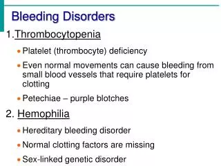
Inherited Bleeding Disorders • A group of inherited diseases • Abnormal and prolonged bleeding - Haemophilia: Coagulation defect, deficiency of Factor VIII/IX - VWD: Defect in platelet adhesion, deficiency of Von Willebrand Factor - Rare coagulation factor deficiencies
How Blood Clots • Coagulation System makes • Fibrin • Forms a supporting network • around the platelet plug • Strengthens the clot • Seals the damaged area of the • blood vessel • Prevents further blood loss
Coagulation System is a • series of linked enzyme • reactions • Enzymes are proteins • called Clotting Factors • Each Factor given a • number • (Roman Numerals) • Factor VIII and Factor IX • are two of the most • important clotting factors
Inheritance of Haemophilia • Inheritance is X-Linked - males affected - females may be carriers • Haemophilia A: ~1/10,000 males • Haemophilia B: ~1/30,000 males
Haemophilia Inheritance XY XX XY XY XX XX Mother is a Carrier: 50% of sons have Haemophilia 50% of daughters are Carriers Important to identify female carriers for Genetic Counselling
Haemophilia A • Lack of Factor VIII in plasma • Range of clinical severity: Severe/moderate/mild • Severity related to Factor VIII activity (FVIII:C) - Severe: 0-2% - Moderate: 2-5% - Mild: 5-30%
Haemophilia A • Lack of Factor VIII in plasma is • caused by mutations in the gene • that makes the Factor VIII protein • F8 gene is located at the tip of the • X chromosome
What is a Gene? • A piece of DNA that contains all the information needed to make a protein • Human Beings have 35,000 genes. • Genes are spread out along the chromosomes • Each gene makes a different protein • Proteins do lots of important jobs in the cell
DNA • Linear molecule, very long • Double Helix • A, G, C, T (bases) • DNA Sequence contains information needed to make a protein
Proteins • Do all the jobs that need done in the cell • Proteins are strings of amino acids • 30 different amino acids • Each amino acid encoded by 3 DNA bases (codon) AGC = Arginine TTC = Phenylalanine GTC = Valine
F8 Gene • F8 Gene is very large – 186,000 bases • Gene is split into Exons and Introns • The 26 exons encode the 2332 amino acid • sequence of the Factor VIII protein • The 26 exons cover 8,000 bases. • Haemophilia A is caused by a mutation of just one base
Mutation Screening • Males with Haemophilia are routinely screened to identify their F8 mutation • Two Purposes: 1. Confirm the diagnosis and the severity 2. Allows screening of related females who may be carriers and at risk of having an affected male child
Identifying F8 Mutations 1. Obtain blood sample from male with Haemophilia 2. Extract DNA sample 3. Isolate his F8 Gene (PCR) 5. Compare patient and normal sequence 6. Look for a difference (Mutation) 4. Determine the DNA sequence of his F8 Gene
What is a Mutation? • A change in the DNA sequence of a gene that causes an inherited disease • Two types of mutation: • Point mutations: a single base is changed • Gross mutation: a change involving a large piece of DNA sequence (eg: deletion mutation)
Point Mutations • Three types of Point Mutation cause Haemophilia: 1. Missense Mutations 2. Nonsense Mutations 3. Frameshift Mutations
Missense Mutations • Missense mutation: base change alters the amino acid encoded at that point G>A mutation: AGC>AAC: Serine>Asparagine C>T mutation: CGT>TGT: Arginine>Cysteine • Changing the amino acid alters the biological activity of the protein • Usually associated with mild Haemophilia
Nonsense Mutations 2. Nonsense Mutations • 64 codons encode 30 amino acids • Three codons act as a signal to indicate the end of the gene during protein synthesis • STOP codons: TGA, TAA and TAG • A nonsense mutation is a base change that creates a new STOP codon C>T: CGA>TGA: Arginine>STOP C>A: TCA>TAA: Serine >STOP
Nonsense Mutations • Nonsense mutations create a new STOP codon in the middle of the gene • Protein synthesis stops too early • A shortened protein is produced (inactive) • Usually causes severe Haemophilia Start Codon Stop Codon
Frameshift Mutations 3. Frameshift mutations: • Caused by the insertion or deletion of a base • Changes the order of the codons in the gene (reading frame) ATT CAG GCA GAA ATA GTA TTT AGA GGG I Q A E I V F R G ATT CAG GTC AGA AAT AGT ATT TAG AGG G I Q V R N V I STOP
Frameshift Mutations • A different set of amino acids is read after the inserted (or deleted) base • Biological activity of the protein is lost • Usually causes severe Haemophilia
Missense Mutation F8 Exon 19 G to A point mutation at nucleotide 6089 Changes Serine 2030 to Asparagine Serine 2030 is highly conserved
Sister of patient is heterozygous carrier of the same mutation Carrier Haemophilia A
Nonsense Mutation F8 Exon 24 C to T Point mutation at nucleotide 6682 Changes Arginine 2228 (CGA) to a STOP codon (TGA) Premature termination of translation gives shortened protein (unstable) Patient has Severe Haemophilia A
Patient’s sister is heterozygous carrier of same mutation Carrier Severe Haemophilia A
Frameshift Mutation Single base (A) deleted at nucleotide 3637 (8 A’s instead of 9) Reading frame changes at codon 1213 Normal sequence: ATT CAG GAA GAA ATA GAA I Q E E I E Mutated sequence: TTC AGG AAG AAA TAG F R K K STOP
Patient’s sister is heterozygous carrier of the same mutation. Deletion of a single base on one allele causes sequences to be out of phase.
60% have 1 of 3 F8 mutations
Von Willebrand Disease First Described by the Finnish Physician, Dr Eric von Willebrand in 1926
In 1926, Erik von Willebrand published an article describing a bleeding disorder he had first observed in some members of a family from the Aland islands The index case was a 5-year old girl who he documented as having a series of life-threatening bleeding episodes. At the age of 14 years, she subsequently bled to death during her fourth menstrual period. Von Willebrand subsequently studied 66 members of the same family and found that 23 of them had symptoms of the same type.
Von Willebrand Disease • Most common IBD in Humans • Variable and mostly mild bleeding tendency • Inherited deficiency of Von Willebrand Factor (VWF) • Caused by VWF gene mutations (usually)
Inheritance of VWD • Inheritance different from • Haemophilia • Males and Females • affected • Autosomal Dominant: only • one defective gene needs to • be inherited for a person to • have the disease • 50% of children of a parent • with VWD will have VWD • themselves VWF/VWF VWF/VWF VWF/ VWF VWF/VWF VWF/VWF VWF/VWF
Bleeding Symptoms in VWD • Distinct from haemophilia • Skin and mucous membranes affected: easy bruising, epistaxis, menorrhagia, excessive bleeding from minor wounds, dental extractions, surgery, childbirth, oral bleeding, GI bleeding • Unlike haemophilia, musculoskeletal bleeding (haemarthroses, muscle haematomas) is rare • Serious bleeding only for severe forms (type 3 VWD).
Von Willebrand Factor • VWF is a large plasma protein • Binding sites for FVIII, collagen and platelets • Important role in blood clotting • Mediates platelet adhesion and aggregation
VWF Multimers • VWF synthesised in endothelial • cells and megakaryocytes • Exists as a series of polymer • chains called Multimers • Multimer structure is important • for role of VWF in blood clotting • VWF stored in granules in • platelets and endothelial cells • VWF multimers released into • plasma in response to blood • vessel injury
Vessel Damage exposes subendothelium to blood • VWF binds to collagen • Gp1b binding site on VWF becomes exposed • VWF binds platelets via Gp1b • Platelets adhere to damaged area
VWF Protects Factor VIII • VWF binds Factor VIII in plasma • Protects FVIII from degradation • No VWF binding: Factor VIII has short half life
How Common is VWD? • Plasma VWF ref range: 50-200 IU/mL • 1% of the population have a level <50IU/mL • Clinically significant VWD only seen in 1:8,000 (0.125%)
VWF and Bleeding Risk As VWF levels get lower, the risk of bleeding increases but relationship is not strong until the VWF level is very low Mild bleeding is common in healthy population and may be due to factors other than VWF level Diagnosis of VWD cannot be made only on the basis of low plasma VWF levels
Plasma VWF levels Vary Widely • Levels affected by genetic and acquired factors • Blood group is major genetic influence. Group O 25% lower the Non-O • Acquired factors: Age, Exercise, Menstrual Cycle, Thyroid Function • Levels increase with age: 6%/decade after 40
Diagnosis of VWD • Three Criteria: • Laboratory tests indicating a deficiency of VWF PLUS • A personal bleeding history • A family history of bleeding
Lab Tests for VWF • VWF: Ag - Measures the amount of VWF protein in plasma • Immunoassay • VWF: RiCoF • Measures VWF activity in plasma
VWF:RiCoF Test • Ristocetin is an obsolete antibiotic • Ristocetin binds to VWF and exposes Gp1b binding site • VWF can bind platelets spontaneously when ristocetin is present • Platelets + ristocetin + Patient plasma • Platelet aggregation measured (platelet aggregometer) • Aggregation is a measure of VWF activity
3 types of VWD exist • Type 1 : Reduced amount of VWF in plasma VWF:Ag and VWF:RCo reduced in parallel • Type 2 VWD: Reduced plasma VWF activity VWF:Ag > VWF:RCo (Ratio >2) • Type 3 VWD: rare, severe form VWF:Ag and VWF:RCo ~0%
Type 2 VWD • Type 2A: Loss of the biggest (HMW) multimers, reduced platelet adhesion • Type 2B: VWF binds to platelets spontaneously • Type 2M: Loss of VWF ability to bind platelets • Type 2N: Defective Factor VIII binding
Type 1 VWD • Reduced plasma VWF levels • Two categories of patients: 1. VWF <20 IU/mL associated with: • VWF gene mutation • Significant bleeding • Strong family history of bleeding • 75% of mutations are missense • Mutations cause secretion failure or rapid clearance from blood
Type 1 VWD 2. VWF levels of 30-50 IU/mL - Up to 1% of population • Less than 50% have a VWF gene mutation • Personal and family bleeding history are less convincing • VWF levels alone are not sufficient for a diagnosis of VWD • Levels should be seen as a modest risk factor for bleeding
Summary • VWD is the most common IBD in humans • Results in a mostly mild bleeding tendency that affects males and females • Diagnosis requires an assessment of VWF levels in conjunction with a personal and family bleeding history • DNA testing is a useful tool to confirm diagnosis