Download
1 / 4
60 likes | 1.03k Vues
VERRUCOUS CARCINOMA DEVELOPED ON A CHRONIC VENOUS ULCER Anton Mihai Țilea , Doru Aurel Chiriță Central Universitary Military Hospital “ Dr. Carol Davila ” Bucharest. Introduction Verrucous carcinoma is a form of well-differentiated squamous cell carcinoma.

E N D
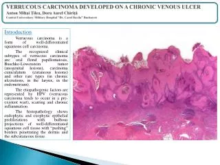
VERRUCOUS CARCINOMA DEVELOPED ON A CHRONIC VENOUS ULCER Anton Mihai Țilea, Doru Aurel ChirițăCentral Universitary Military Hospital “Dr. Carol Davila” Bucharest Introduction Verrucouscarcinoma is a form of well-differentiated squamous cell carcinoma. The recognized clinical subtypes of verrucous carcinoma are: oral florid papillomatosis, Buschke-Löwensteintumor(anogenital lesions), carcinoma cuniculatum (cutaneous lesions) and other rare types (in chronic ulcerations, in the larynx, in the endometrium). The etiopathogenic factors are represented by HPV (verrucous carcinoma tends to occur in a pre-existent wart), scarring and chronic inflammation. The histopathology shows endophytic and exophytic epithelial proliferations with bulbous projections of well-differentiated squamous cell tissue with “pushing” borders penetrating the dermis and the subcutaneous tissue.
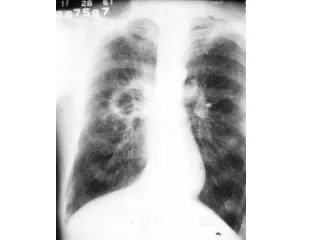
VERRUCOUS CARCINOMA DEVELOPED ON A CHRONIC VENOUS ULCER Anton Mihai Țilea, Doru Aurel ChirițăCentral Universitary Military Hospital “Dr. Carol Davila” Bucharest Case presentation We present the case of a 60 years old patient with a personal history of chronic venous insufficiency with multiple surgeries performed on the right calf for varicose veins. The patient presented to our clinic for a malodorous ulcer covering almost one third of the right calf, in some places almost circumferential, with relatively well-defined borders, the base being covered with serohematic debris. Some papillomatous, warty lesions were present on the surface. The lesion had a chronic evolution of about 2 years.
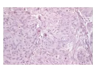
VERRUCOUS CARCINOMA DEVELOPED ON A CHRONIC VENOUS ULCER Anton Mihai Țilea, Doru Aurel ChirițăCentral Universitary Military Hospital “Dr. Carol Davila” Bucharest Histopathology Thebiopsy was taken from a papillomatous, warty lesion. It showed an epithelial proliferation, exophytic with atypical parakeratosis and endophytic with bulbous projections of well-differentiated squamous cell tissue with “pushing” borders penetrating deep into the reticular dermis and the subcutaneous tissue.
VERRUCOUS CARCINOMA DEVELOPED ON A CHRONIC VENOUS ULCER Anton Mihai Țilea, Doru Aurel ChirițăCentral Universitary Military Hospital “Dr. Carol Davila” Bucharest Treatment Systemic and topical antibiotics were used to treat the local infection. Extensive surgery was performed with the excision of the entire ulcerative lesion and the primary defect was repaired by skin grafting. Post-grafting the lesion healed well. Conclusions • The biopsy was essential as it allowed the patient to receive the adequate surgical therapy and subsequently to have the ulcerative lesion healed. • The chronic evolution of a non-responsive ulcerative lesion should determine every clinician to perform a biopsy and perhaps extensive lesions like in this case will be avoided. Bibliography • Eduardo Calonje, Thomas Brenn, Alexander Lazar, Phillip H McKee - McKee’s Pathology of the Skin 4th edition 2012, p1131-1134. • Patricia Senet, MD; Patrick Combemale, MD; Clelia Debure, MD; Nathalie Baudot, MD; Laurent Machet, MD, PhD; Mounir Aout, PhD; Eric Vicaut, MD, PhD; Catherine Lok, MD, PhD; for the Angio-Dermatology Group of the French Society of Dermatology– “Malignancy and Chronic Leg UlcersThe Value of Systematic Wound Biopsies: A Prospective, Multicenter, Cross-sectional Study” - Arch Dermatol. 2012;148(6):704-708. doi:10.1001/archdermatol.2011.3362