Download
1 / 12
120 likes | 376 Vues
SQUAMOUS CELL CARCINOMA ARISING IN A DERMOID CYST OF THE OVARY. Scibetta Nunzia , Marasà Lorenzo. C.O.U. of Pathologic Anatomy, ARNAS-Civico Hospital, Palermo, Italy. Mature cystic teratomas make up almost 20% of all ovarian neoplasms.

E N D
SQUAMOUS CELL CARCINOMA ARISING IN A DERMOID CYST OF THE OVARY. Scibetta Nunzia , Marasà Lorenzo C.O.U. of Pathologic Anatomy, ARNAS-Civico Hospital, Palermo, Italy.
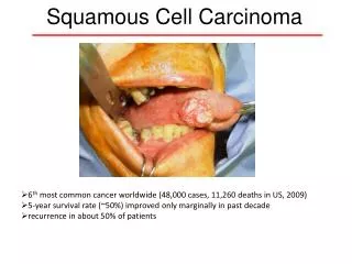
Mature cystic teratomas make up almost 20% of all ovarian neoplasms. Squamous cell carcinoma (SCC) arising in mature cystic teratoma is rare, approximately 1-3%, and affects elderly persons.
We report a case of SCC originating from a dermoid cyst of left ovary in a 78 year-old woman.
Materials and methods The study included a 78 year-oldpatient underwent unilateral salpingo-oophorectomy. The specimens were fixed in 4% formaldehyde and embedded in paraplast. Sections 4 micron thick were stained with H&E and PAS. Immunohistochemistry was performed.
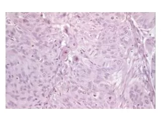
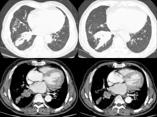
Results The left ovary was cystic, unilocular, mass measuring 10x7x5 cm, with external and internal surfaces pink and smooth and showed abundant hair and yellow sebaceous material. Microscopically, the cystic wall was lined by mature and immature squamous epithelium, involved by extensive SCC in situ, distinguished by a diffuse expansion of epithelium by large cells with a variety of atypical changes, such as cytoplasmic vacuolization, nuclear hypercromasia, marked anaplasia.
Results The tumor cells were positive for CKAE3, EMA, CEA, ulex europaeus, negative for S100, vimentin. Microscopic invasion of the wall by single malignant cells and small clusters was observed. No malignant cells were observed in peritoneal washing. The patient was categorized as FIGO stage IA. She is now doing well without recurrence of disease six months after the surgery .
A B Figs. A, B, C The cystic wall is lined by mature and immature squamous epithelium (H&E, x100) C
Epidermal expansion with marked anaplasia and nuclear hypercromasia are evident (H&E x 200)
Microscopic invasion of the wall by small cluster of malignant cells (H&E x200)
Corpus albicans in the wall of mature cystic teratoma (H&E, x 100)
Conclusions SCC originating from dermoid cysts are rare tumors. Every case may not have the same clinico-pathologic characteristics and management should be individualized. To our knowledge, it is the first report of a extensive SCC in situ with microinvasion arising in a mature cystic ovarian teratoma.
References • Basgul A, Gokaslan H, Kavak ZN, Guducu N, Eren F : Squamous cell carcinoma developing in a huge dermoid cyst of the ovary in an 80-year-old woman : case report . Eur J Gynecol Oncol 2007, 28: 63-66. • Hurwitz JL, Fenton A, McCluggage WG, McKenna S : Squamous cell carcinoma arising in a dermoid cyst of the ovary: a case series . BJOG 2007, 114 : 1283-1287. • Powell JL, Stinson JA, Connore GP, Shiro BS, Mattison M : Squamous cell carcinoma arising in a dermoid cyst of the ovary. Gynecol Oncol. 2003, 89 : 526-528.