Download
1 / 8
80 likes | 314 Vues
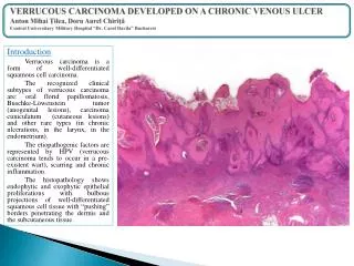
Verrucous carcinoma of the foot. Deba P Sarma , MD Omaha. M 63, left foot. Markedly hyperkeratotic and centrally ulcerated lesion measuring 3.0 x 1.7 on the plantar surface.

E N D
Verrucous carcinoma of the foot Deba P Sarma, MD Omaha
M 63, left foot Markedly hyperkeratotic and centrally ulcerated lesion measuring 3.0 x 1.7 on the plantar surface
Marked hyperkeratosis, papillomatosis, acanthosis, irregular elongation of the rete ridges with pushing type of invasion with bulbous proliferation of well-differentiated keratinocytes
Burrowing invasion of the dermis by bulbous broad columns of well differentiated squamous epithelium
Diagnosis: -Verrucous carcinoma. Verrucous carcinoma is an uncommon, well-differentiated variant of squamous cell carcinoma that is regarded as a low grade locally invasive malignant tumor with rare potential for distant metastasis. Verrucous carcinoma is usually seen in oral cavity, genitoanal region, and plantar surface of foot. They are known to arise in chronic ulcers, in skin after radiation therapy, old scars, and rarely in the amputation stump. Verrucous carcinoma is a low-grade squamous cell carcinoma that can be classified into three major groups based on the sites of origin: verrucous carcinoma of oral cavity, verrucous carcinoma of genitoanal region, and plantar verrucous carcinoma.
Plantar verrucous carcinoma, also called epithelioma cuniculatum, was first described by Aird in 1954. The lesion initially shows a striking resemblance to an intractable plantar wart. As the exophytic mass grows, it shows a great tendency toward deep, penetrating growth, resulting in numerous deep crypts resembling the burrows of rabbits, hence the name cuniculatum. Although the tumor grows slowly, it will eventually invade the plantar fascia, may destroy metatarsal bones, and may invade the skin of the dorsum of the foot.
Histological features favoring the diagnosis of verrucous carcinoma include: (1) deep invasion, (2) branching tunnels and keratin-filled clefts, (3) large pale keratinocytes with large nuclei, and (4) edematous stroma with chronic inflammatory cells. Histolologic differentiation from pseudoepitheliomatous hyperplasia or benign verrucous hyperplasia remains a difficult problem. Clinical history of non-healing verrucous lesion over a long period along with the histologic finding of bulbous pushing-type of invasion by pale well-differentiated keratinocytes in the dermis will be highly suggestive of a verrucous carcinoma.
Ref: Sarma DP, Wang JF, Bewtra C, Lee LMJ (2007). Verrucous carcinoma arising in a chronic non-healing ulcer of the foot of a diabetic patient. The Internet J Dermatol, 5(1). Indexed by Google Scholar.