Download
1 / 20
800 likes | 2.85k Vues
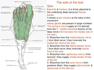
Sole of the foot. Superficial fascia Deep fascia Cutaneous nerve Medial calcanean branches of tibial n Lateral planter n Medial planter n. Sole of the foot. Plantar Aponeurosis Apex Base Function fixes the skin of the sole Protects the deeper str

E N D
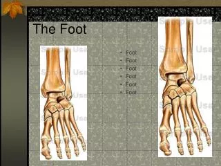
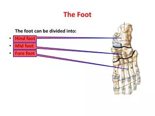
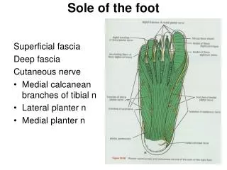
Sole of the foot Superficial fascia Deep fascia Cutaneous nerve • Medial calcanean branches of tibial n • Lateral planter n • Medial planter n
Sole of the foot Plantar Aponeurosis Apex Base Function • fixes the skin of the sole • Protects the deeper str • Maintain the longitudinal arches of the foot • Origin of the muscles of the first layer of the sole
Sole of the foot Muscles of the first layer Flexor digitorum brevis- Origin- Medial tubercle of the calcaneum, plantar aponeurosis and medial and lateral intermuscular septa Insertion-4 tendons for 4 lateral toes Nerve supply-medial planter nerve Action- flexion of the toes at the proximal interphalangeal jts and metatarsophalangeal jts
Sole of the foot Abductor hallucis- Origin: medial tubercle of calcaneum, flexor retinaculum, deep fascia covering and medial intermuscular septum Insertion-medial side of the base of the proximal phalanx of the great toe Nerve supply-medial plantar nerve Action- Abduction of the great toe away from the second toe
Sole of the foot Abductor digiti minimi Origin-medial and lateral tubercles of the calcaneum, lateral intermuscular septum and deep fascia covering it Insertion-lateral side of the base of the proximal phalanx of the little toe Nerve supply- main trunk of the lateral plantar nerve Action- abduction of the little toe
Sole of the foot Muscles of the first layer of the sole
Sole of the foot Muscles of the second layer Flexor digitorum longus- Origin: upper 2/3rd of the medial part of the post surface of the tibia below the soleal line Insertion: 4 tendons each into the planter surface of the distal phalanx of 2nd to 5th toes Nerve supply- tibial nerve Action- Plantar flexion of the lateral four toes, planter flexion of the ankle jt and maintains medial longitudinal arch
Sole of the foot Flexor digitorum accessorius Origin: medial head from the medial concave surface of the calcaneum and from its medial tubercle and lateral head in front of the lateral tubercle and from the long planter ligament Insertion- lateral side of the tendon of the flexor digitorum longus Nerve supply-Main trunk of the lateral plantar nerve Action- striaghtens the pull of the long flexor tendons and flexes the toes through the long tendons
Sole of the foot Lumbricals Origin: long flexor digitorum longus Insertion:medial sides of the metatarsophalangeal jts of the lateral toes and then dorsally for the insertion into the extensor expansion Nerve supply- 1st by the medial plantar n and other 3 by the deep branch of the lateral plantar nerve Action- maintain extension of the digits at the interphalangeal jts ie in walking and running the toes do not buckle under
Sole of the foot Flexor hallucis longus Origin: lower 3/4th of the post surface of the fibula and interosseous membrane Insertion: plantar surface of the base of the distal phalanx of the great toe Nerve supply-tibial nerve Action-plantar flexor of the big toe, plantar flexion of the ankle jt and maintains the medial longitudinal arch
Sole of the foot Muscles of the third layer of the sole Flexor hallucis brevis- Origin: Y shaped, lateral limb from the medial part of the plantar surface of the cuboid bone, behind the groove for the peroneus longus and from the adjacent side of the lateral cuneiform bone and medial limb direct continuation of the tendon of the tibialis posterior into the foot Insertion:2slips into lateral and medial parts, ends into corresponding side of the base of the proximal phalanx of the great toe Nerve supply-medial plantar nerve Action- Flexes the proximal phalanx at the metatarsophalangeal jt of great toe
Sole of the foot Adductor hallucis origin: oblique head-base of the 2nd,3rd and 4th metatarsals, from the sheath of the tendon of the peroneus longus and transverse head-deep metatarsal lig and the plantar lig of the metatarsophalangeal jts of 3rd, 4th and 5th toes Insertion: lateral side of the base of the proximal phalanx of the big toe, in common with the lateral tendon of the flexor hallucis longus Nerve supply-deep branch of lateral plantar nerve Action- Adductor of the great toe towards the second toe and maintains transverse arch of the foot
Sole of the foot Flexor digiti minimi brevis Origin: base of the 5th metatarsal bone and sheath of the tendon of the peroneus longus Insertion: lateral side of the base of the proximal phalanx of the little toe Nerve supply-superficial branch of the lateral plantar n Action-flexes the proximal phalanx at the metatarsophalangeal jt of the little toe
Sole of the foot Muscles of the fourth layer of the sole Interosseous muscles • 3 plantar interossei • 4 dorsal interossei Action-plantar interossei are adductors of the 3rd 4th and 5th toes and the dorsal interossei are abductors.
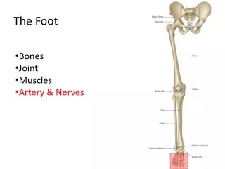
Sole of the foot Plantar vessels • Medial • Lateral
Sole of the foot Nerves • Lateral plantar nerve
Sole of the foot Nerve Medial plantar nerve
Sole of the foot Plantar arch Branches- • 4 plantar metatarsal arteries • 3 proximal perforating arteries • The distal ends of the plantar metatarsal arteries gives off a distal perforating art which joins with distal part of the corresponding dorsal metatarsal artery