Download
1 / 16
170 likes | 215 Vues
Discover the intricate details of the sole of the foot, including its skin, fascia, and muscles, along with nerve supplies and functions. Learn about the fibrous bands, sensory nerve branches, and the significance of the plantar aponeurosis.

E N D
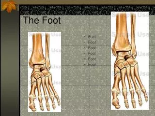
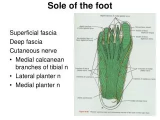
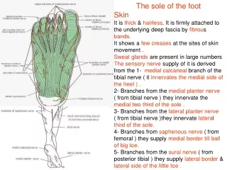
The sole of the foot Skin It is thick & hairless. It is firmly attached to the underlying deep fascia by fibrous bands. It shows a few creases at the sites of skin movement . Sweat glands are present in large numbers The sensory nerve supply of it is derived from the 1- medial calcaneal branch of the tibial nerve ( it innervates the medial side of the heel ) . 2- Branches from the medial planter nerve ( from tibial nerve ) they innervate the medial two third of the sole . 3- Branches from the lateral planter nerve ( from tibial nerve )they innervate lateral third of the sole. 4- Branches from saphenous nerve ( from femoral ) they supply medial border till ballof big toe. 5- Branches from the sural nerve ( from posterior tibial ) they supply lateral border & lateral side of the little toe .
Deep fascia The plantar aponeurosis is triangular and occupies the central area of the sole . It is formed as a thickening of the deep fascia . Its apex is attached to the medial &lateral tubercles of the calcaneum. Its base is divided into 5 slips at the bases of the toes .Each slip divides into 2 bands , one passing superficially to the skin and the other passing deeply to the root of the toe. Then each deep band divides into 2 bands which diverge around the flexor tendons and fuse with the fibrous flexor sheath & the deep transverse ligament . The deep fascia which covers the abductor of the big & little toes is thinner & weak . At the junction of the medial & lateral borders of plantar aponeurosis with the thinner deep fascia fibrous septa pass superiorly to form fascial spaces of the sole. The function of the plantar aponeurosis 1- give firm attachment to the overlying skin 2- protect vessels, nerves, tendons & synovial sheaths . 3- assist in maintaining the arches of the foot
First layer of the plantar muscles of the right foot 1- Abductor hallucis medial plantarnerve 2- Flexor digitorum brevis medial planter nerve 3- Abductor digiti minimi lateral planter nerve
Second layer of the plantar muscles of the right foot 1- Quadratusplantae lateral planter nerve 2- Lumbricals first …….medial planter nerve the remainder 3 …lateral planter n 3- Flexor digitorum longus tendon tibial nerve 4- Flexor hallucis longus tendon tibial nerve
Third layer of the plantar muscles of the right foot 1- flexor hallucis brevis medial plantar nerve 2- Adductor hallucis deep branch of the lateral plantar n 3- flexor digiti minimi brevis lateral plantar nerve
Fourth layer of the plantar muscles of the right foot 1- Dorsal interossei lateral plantar nerve 2- Plantar interossei lateral plantar nerve 3- Peroneus longus tendon superficial peroneal nerve 4- Tibialis posterior tendon tibial nerve
Tendons of the second layers 1- flexor hallucis longus : Its tendon enters the sole by passing behind the medial malleolusbeneath the flexor retinaculum . It runs forward below the sustentaculum tali and crosses deep to the flexor digitorum longus tendon. It then enters the fibrous sheath of the big toe to insert into the base of the distal phalanx. 2- flexor digitorum longus : Its tendon enters the sole by passing behind the medial malleolusbeneath the flexor retinaculum. It passes forward along the medial surface of the sustentaculum tali then crosses the tendon of FHL. It receives on its lateral border the insertion of the quadratusplantae muscle. It divides into 4 tendons giving origin to lumbricalmuscles . They enter the fibrous sheath of the lateral 4 toes. Each tendon perforates the corresponding tendon of FDB& inserted into the base of the distal phalanx .
Long tendons of the 4th layer 1-Tibialis posterior : It enters the foot from behind the medial malleolus . It passes beneath the flexor retinaculum and runs downward & forward above the the sustentaculum tali to be inserted mainly into the tuberosity of the navicular. Small tendinous slips pass to the cuboit, cuneiforms and to the bases of 2nd, 3rd, and 4th MT 2- Peroneus longus : It enters the foot behind the lateral malleolus. It passes beneath the superior & inferior peroneal retinachla & lies below the peroneal tubercle on the lateral side of the calcaneum. It enters a groove on the inferior aspect of the cuboid bone It runs obliquely to insert into the base of the first metatarsal & adjacent part of the medial cuneiform . It held by a band from the long plantar ligament .
Fibrous flexor sheaths (deep fascia ) The inferior surface of each toe, from the head of the metatarsal to base of the distal phalanx , is provided with strong fibrous sheath, which is attached to the sides of the phalanges. Its distal end is closed & attached to the base of the distal phalanx. It with the inferior surface of the phalanges & the interphalangeal joints form a blind tunnel in which lie the flexor tendons of the toe. It is thick over the phalanges but thin and lax over the joints. The proximal ends of the fibrous sheaths of the toes receive the deeper parts of the 5 slips of the plantar aponeurosis.
Synovial flexor sheaths 1- The tendon of the flexor hallucis longus is surrounded by a synovial sheath , which extends upward behind the medial malleolus & above the flexor retinaculum. Distally it extends to the base of the 1st metatarsal . As it enters the fibrous sheath it acquires a digital synovial sheath . 2- The flexor digitorum longus synovial sheath extends upward behind the medial malleolus & for a short distance above the flexor retinaculum. Distally it extends to the navicular.As the 4 tendons enter the fibrous flexor sheath,it acquires a digital synovial sheath . 3- The tibialis posterior synovial sheath passes beneath the flexor retinaculum to the tuberosity of the navicular bone .
4- The tendon of the peroneus longus is surrounded by a synovial sheath . It passes beneath the peroneal retinacula . As the tendon winds around the lateral margin of the cuboid . It is thickened & contains a sesamoid cartilage. A second synovial sheath surrounds the tendon as it crosses the sole.
Arteries of the sole 1- Medial plantar artery It is the smaller branch of the terminal branches of the posterior tibial artery. It arises beneath the flexor retinaculum and passes forward deep to the abductor hallucis muscle . It ends by supplying the medial side of the big toe . During its course it gives off numerous muscular , cutaneous and articular branches. Veins of the soleMedial & lateral plantar veins unite behind the medial malleolus to form the posterior tibial venae comitantes .
2- Lateral plantar artery It is the larger branch of the terminal branches of the posterior tibial artery . It arises beneath the flexor retinaculum and passes forward deep to the abductor hallucis & the flexor digitorum brevis . On reaching the base of the 5th metatarsal, it is under the cover of abductor digiti minimi . It curves medially to form the plantar arch and at the proximal end of the 1st intermetatarsal space joins the dorsalis pedis artery . During its course , it gives off numerous muscular, cutaneous and articular branches . The plantar arch gives off plantar digital arteries to the toes .
Nerves of the sole 1- Medial plantar nerve It is a terminal branch of the tibial nerve . It arises beneath the flexor retinaculum . It runs forward deep to the abductor hallucis with the medial plantar nerve. It comes to lie in the interval between the abductor hallucis and the flexor digitorum brevis. Branches : 1- muscular to abductor hallucis, flexor digitorum brevis, flexor hallucis brevis and the 1st lumbrical muscle . 2- cutaneous : plantar digital nerves which run to the sides of the medial 3 and half toes. They extend onto the dorsum and supply the nail beds & tips of the toes.
Lateral plantar nerve It is a terminal branch of the tibial nerve . It arises beneath the flexor retinaculum . It runs forward deep to the abductor hallucis & the flexor digitorum brevis with the lateral plantar artery. On reaching the base of the 5th metatarsal , it divides into superficial & deep branches . Branches : 1- From the main trunk : cutaneous branches to the skin of the lateral part of the sole and muscular to quadratus plantae ( 2nd ) & abductor digiti minimi.
2- From the superficial terminal branch : To the flexor digiti minimi & the interosseous muscles of the 4th intermetatarsal space. Plantar digital branches pass to the sides of the lateral one and half toes . They extend onto the dorsum and supply the nail beds & tips of the toes. 3- From the deep terminal branch : It curves medially with the lateral plantar artery (plantar arch ). It supplies the adductor hallucis ; the 2nd , 3rd and 4th lumbricals ; all the interossei except those of the 4th intermetatarsal space .