Download
1 / 37
410 likes | 890 Vues
The Sole of the Foot. (the planter aspect of the foot). Dr. Zeenat Zaidi. Skin. Thick & hairless , lacks pigmentation Possesses abundant sweat glands Firmly bound down to the underlying deep fascia by numerous fibrous bands Shows few flexor creases at the sites of skin movement.

E N D
The Sole of the Foot (the planter aspect of the foot) Dr. ZeenatZaidi
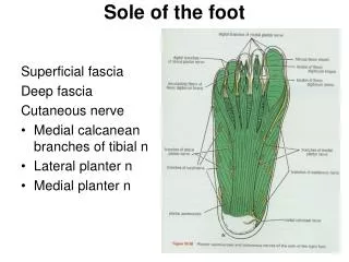
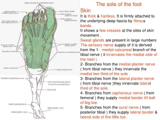
Skin • Thick & hairless, lacks pigmentation • Possesses abundant sweat glands • Firmly bound down to the underlying deep fascia by numerous fibrous bands • Shows few flexor creases at the sites of skin movement. • The subcutaneous tissue contains a lot of fat, especially in the heel • Extremely sensitive to touch due to a high concentration of nerve endings. This makes the sole sensitive to surfaces that are walked on
Cutaneous Nerve Supply • Medial side of the heel: • Medial calcaneal branch of the tibial nerve • Medial 2/3 of the sole: • Branches from the medial plantar nerve • Lateral 1/3 of the sole: • Branches from the lateral plantar nerve • Along the Medial border: • Saphenous nerve • Along the Lateral border: • Sural nerve
Deep Fascia • Lies beneath the subcutaneous tissue andsurrounds the intrinsic foot muscles • Much thicker in the central part and thinner where it covers the intrinsic muscles of big toe and little toe • The central thicker part forms triangular planter aponeurosis
Planter Aponeurosis • A triangular thickening of the deep fascia that protects the underlying nerves, blood vessels, and muscles. • Apex is attached to the medial and lateral tubercles of the calcaneum. • Base divides into five slips that pass into the toe • Each slip further divides into the: • Superficial band to the skin • Deep band passing to the root of the toes, where it divides into two, diverging along the flexor tendons and fusing with the fibrous sheath and the deep transverse ligaments
From each of these borders, fibrous septa pass superiorly into the sole and take part in the formation of the fascial spaces of the sole. The medial and lateral borders of the thick aponeurosis are continuous with the thinner deep fascia covering the abductors of the big and little toes.
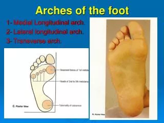
Functions • To give a firm attachment to the overlying skin • To protect the underlying vessels, nerves and tendons and their synovial sheaths • To assist in maintaining the arches of the foot.
Muscles of the Sole • The sole contains both Extrisic & Intrinsic muscles • These muscles: • Help to flex, extend, abduct, and adduct the toes • Enable the toes to lift and curl. • Support the arches of the foot • Are supplied by branches of tibial nerve • Are supplied by branches of posterior tibial artery • Are arranged in four layers.
From superficial to deep • The muscles of the first layer are: • Abductor hallucis • Flexor digitorumbrevis • Abductor digitiminimi
The muscles of the second layerare: Accessory flexor (quadratusplantae) Lumbricals Tendons of the flexor digitorumlongusfrom which the lumbricals arise
The muscles of the third layer are: • Flexor hallucisbrevis • Adductor hallucis • oblique head • transverse head • Flexor digitiminimibrevis
The muscles of the fourth layer are: • Dorsal interossei • Plantar interossei • Tendon of the peroneuslongus • Tendon of the tibialis posterior
Long Tendons of the Sole of the Foot • Flexor DigitorumLongus Tendon • Enters the sole by passing: • behind the medial malleolus • beneath the flexor retinaculum. • Passes forward across the medial surface of the sustentaculumtali • Then crosses the tendon of flexor hallucislongus, from which it receives a strong slip. • It is here that it receives on its lateral border the insertion of the quadratusplantaemuscle.
Each tendon perforates the corresponding tendon of flexor digitorumbrevis and passes on to be inserted into the base of the distal phalanx (compare with the insertion of the flexor digitorumprofundus in the hand) The tendon now divides into its four tendons of insertion, which pass forward, giving origin to the lumbrical muscles. The tendons then enter the fibrous sheaths of the lateral four toes
Flexor HallucisLongus Tendon • Enters the sole by passing: • behind the medial malleolus • beneath the flexor retinaculum. • It runs forward below the sustentaculumtaliand crosses deep to the flexor digitorumlongus tendon, to which it gives a strong slip. • It then enters the fibrous sheath of the big toe and is inserted into the base of the distal phalanx.
Fibrous Flexor Sheaths • The inferior surface of each toe (from the head of the metatarsal bone to the base of the distal phalanx), is provided with a strong fibrous sheath, which is attached to the sides of the phalanges. Their proximal ends receive the deeper parts of the slips of plantar aponeurosis Their distal ends are closed and are attached to the base of the distal phalanges
Synovial sheath Fibrous sheath Flexor tendon Phalanx The fibrous sheath, together with the inferior surfaces of the phalanges and the interphalangeal joints, forms a blind tunnel for the flexor tendons of the toes
Synovial Flexor Sheaths • The tendons of the flexor muscles are surrounded by synovial sheaths • The synovial sheath of the flexor hallucislongus extends: • Upwards behind the medial malleolusabove the flexor retinaculum • Ends distally at the base of the first metatarsal bone. • The synovial sheaths of flexor digitorumlongusextend: • Above the flexor retinaculum • Extend distally as far as the navicular bone
PeroneusLongus Tendon Enters the foot from behind the lateral malleolus Runs obliquely across the sole to be inserted into the base of the first metatarsal bone and the adjacent part of the medial cuneiform. The tendon grooves the inferior surface of the cuboidwhere it is held in position by the long plantar ligament and is surrounded by a synovial sheath
Tibialis Posterior Tendon • Enters the foot from: • behind the medial malleolus. • beneath the flexor retinaculum • Runs downward and forward above the sustentaculumtalito be • Inserted mainly into the tuberosity of the navicular. • Small tendinous slips pass to the cuboid and the cuneiforms and to the bases of the second, third, and fourth metatarsals. • The tendon is surrounded by a synovial sheath.
Arteries of the Sole of the Foot • Posterior tibial artery enters the foot: • Medially under the medial malleolus • Deeper to flexor retinaculum • Divides to give the medial and lateral plantar arteries which supply the sole
Medial Plantar Artery • The smaller of the terminal branches of the posterior tibial artery. • Arises beneath the flexor retinaculumand passes forward deep to the abductor hallucis • Ends by supplying the medial side of the big toe Gives numerous muscular, cutaneous & articular branches
Lateral Plantar Artery • The larger of the terminal branches of the posterior tibial artery, arises beneath the flexor retinaculum • Passes forward deep to the abductor hallucisand the flexor digitorumbrevis • On reaching the base of the fifth metatarsal bone, the artery curves medially to form the plantar arch • At the proximal end of the first intermetatarsal space joins the dorsalispedis artery Gives numerous muscular, cutaneous & articularbranches and plantar digitalarteries to the toes.
Planter Arch • Formed by lateral plantar artery • Anastomoses with the dorsal pedis arteryby way of a perforating artery which pierces through the proximal end of the first intermetatarsal space • The arch gives rise to several metatarsal branches which split into digital branches.
Veins of the Sole of the Foot • Medial and lateral plantar veins accompany the corresponding arteries, and they unite behind the medial malleolusto form the posterior tibialvenaecomitantes.
Nerves of the Sole of the Foot • Tibial nerveenters the foot medially: • Under the medial malleolus • Deeper to flexor retinaculum • Divides to give the medial and lateral plantar nerves which supply the sole
Medial Plantar Nerve Runs forward deep to the abductor hallucis, with the medial plantar artery Comes to lie in the interval between the abductor hallucisand the flexor digitorumbrevismuscles
Branches • Muscular branches to the: • Abductor hallucis • Flexor digitorumbrevis • Flexor hallucisbrevis • First lumbrical muscle • Cutaneous branches: • Plantar digital nerves run to the sides of the medial 3½ toes • The nerves extend onto the dorsum and supply the nail beds and the tips of the toes.
Lateral Plantar Nerve • Runs forward deep to the abductor hallucis and the flexor digitorumbrevis, in company with the lateral plantar artery • On reaching the base of the fifth metatarsal bone, it divides into: • Superficial • Deep branches
Branches • From the main trunk • Muscular branches to the: • Quadratus plantae • Abductor digiti minimi • Cutaneousbranches to the: • skin of the lateral part of the sole
From the superficial terminal branch • Muscular branches to the: • Flexor digitiminimi • Interosseous muscles of the fourth intermetatarsal space • Cutaneous branches • Plantar digital branches to the sides of the lateral 1½ toes. • Extend onto the dorsum and supply the nail beds and tips of the toes.
From the deep terminal branch • Muscular branches to the: • Adductor hallucis • Second, third, and fourth lumbricals • All the interossei, except those in the fourth intermetatarsal space
Ligaments of the sole of foot CU N N CA CA Long plantar ligament stretches from the calaneumto the cuboid & to the bases of the 2nd, 3rd & 4th metatarsal bones Short planter (plantar calcaneocuboid) ligament, connects the calcaneum to the cuboid, lieson the deep aspect of the long plantar ligament
N CA This ligament is also know as the spring ligament since it is believed to give a spring-like action to the foot when walking. T N CA Plantar calcaneonavicular ligamentextends from the calcaneus to the navicular bone and prevents the head of the talus from pushing down between the calcaneus and the navicular bones.
D Under 2 years: toes are extended • Soles are the site of the planter (Babinski’s) reflex • When the sole of the foot is stroked firmly on the outer side from the heel to the front in persons over the age of 2 years • Normal response is planterflexion (flexion) of the toes. (Negative Babinski's response) • Abnormal response is dorsiflexion of the big toe and often a fanning of the other toes (Positive Babinski's response) • Under 2 years of age, extension of the toes is the normal response