Download

1 / 26
280 likes | 343 Vues
Explore cellular injury, adaptations, and aging processes affecting cells, tissues, organs, and organisms. Discover reversible and irreversible changes, including apoptosis and necrosis. Learn the role of telomeres in aging.

E N D
Cells Tissues Organs Systems Organism
Adaptations Change in size Change in number of cells Change into another type of cell
Atrophy • Decreased size & function • Metabolic processes shut down to conserve energy • Due to • decreased demand • ischemia • lack of nerve or hormonal stimulation • chronic inflammation
Hypertrophy • Increased size & functional capacity • Due to • hormonal stimulation • increased functional demand
Hyperplasia • Increase in number of cells • Due to • hormonal stimulation • increased functional demand • chronic stress or injury
Dysplasia • Disorderly overgrowth of cells • Premalignant • Reversible
Metaplasia • One cell type to another • Reversible
Reversible or Irreversible • Adaptations may be normal physiological responses to stimuli, or pathological conditions Reversible Injury • Functional or morphological changes reverse when stimulus is removed, even if cellular injury has begun: • Moderately reduced oxidative phosphorylation of ATP slows active transport • Aqueous vacuoles may bud from ER—hydropic change • Fatty vacuoles may appear in cytoplasm—fatty change
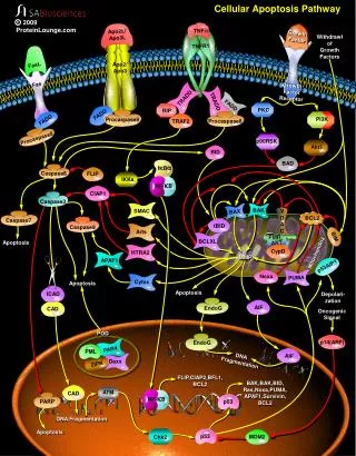
Severe disruption of compartmentalization triggers either necrosis or apoptosis—cells die • Necrosis: swelling, protein denaturation and digestion, membrane breakdown and blebbing • Apoptosis: shrinkage, fragmentation, phagocytosis
Reversible damage – cellular swelling Cellular swelling (synonyms: hydropic change, vacuolar degeneration, cellular edema) is an acute reversible change resulting as a response to nonlethal injuries. It is an intracytoplasmic accumulation of water due to incapacity of the cells to maintain the ionic and fluid homeostasis. It is easy to be observed in parenchymal organs : liver (hepatitis, hypoxia), kidney (shock), myocardium (hypoxia, phosphate intoxication). It may be local or diffuse, affecting the whole organ.
Reversible damage – fatty change Intracellular accumulations of a variety of materials can occur in response to cellular injury. Here is fatty metamorphosis (fatty change) of the liver in which deranged lipoprotein transport from injury (most often alcoholism) leads to accumulation of lipid in the cytoplasm of hepatocytes.
Necrosis • Pathologic cell death • Usually in a collection of cells fed by a single artery
Apoptosis • Programmed cell death • Especially during fetal development • In response to hormonal cycles (e.g. endometrium) • Normal turnover in proliferating tissues (e.g. intestinal epithelium) • Cells shrink, not swell • Nuclei condense and DNA fragments • Cells fragment into membrane-bound bits • Bits are phagocytosed by macrophages
The main factors acting in aging process and the functional relationship between them
Cell cycle is regulated by different specific proteins, cancer supressors, cyclins, and MAP kinases. When these proteins are damaged by mutations cell cycle regulation can be disturbed.
We know genes concerned with pathological aging. When they are damaged organism ages much faster. These genes are named gerontogenes - aging genes. Genetic polymorphisms (determining individual's longevity) are found. The existence of longevity gene is still very real. Some age linked diseases are known in medical practice (Werner's, Bloom's, Cocaine's syndromes, progery and other). Patents had damaged various gerontogenes. It was observed that these genes encoded replication, transcription and repair machinery components of the cell.
Telomeres are the terminal parts of eukaryotic chromosomes. The influence to aging of telomeres is highly discussed. They are called "molecular clock" of the cell. • Cell division times are correlated with telomere length. After each cell division telomeres get shorter. When telomere shortens to the critical stage, the intensity of cell division significantly decreases, and then cell differentiates and ages.
CHROMOSOME TELOMERE 3’ TTAGGGTTAGGGTTAGGGTTAGGGTTAGGG 5’ AATCCCAATCCC
What are telomeres? • Telomeres are… • Repetitive DNA sequences at the ends of all human chromosomes • They contain thousands of repeats of the six-nucleotide sequence, TTAGGG • In humans there are 46 chromosomes and thus 92 telomeres (one at each end) • senescent cells have shorter telomeres • length differs between species • in humans 8-14kb long • telomere replication occurs late in the cell cycle
How Does Telomerase Work? • Telomerase works by adding back telomeric DNA to the ends of chromosomes, thus compensating for the loss of telomeres that normally occurs as cells divide. • Most normal cells do not have this enzyme and thus they lose telomeres with each division.