Download
1 / 102
1.05k likes | 1.61k Vues
Arterial Blood Gas Interpritation. Mark Bromley PGY-2. Why ABGs?. Important clinical info Quick other labs (i.e. faster than a CBC) Fun to interpret. Why Not?. ABG analysis is not without drawbacks. It is painful! Complications Local hematoma

E N D
Arterial Blood Gas Interpritation • Mark Bromley • PGY-2
Why ABGs? • Important clinical info • Quick other labs (i.e. faster than a CBC) • Fun to interpret
Why Not? • ABG analysis is not without drawbacks. • It is painful! • Complications • Local hematoma • Arterial dissection and thrombosis (rarely) • Technically difficult, particularly in children and elderly patients, and several attempts may be required.
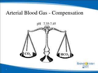
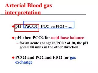
Normal ABG parameters • pH 7.40 • PCO2 40 mmHg • [HCO3] 24 mmol/l • Anion Gap < 12
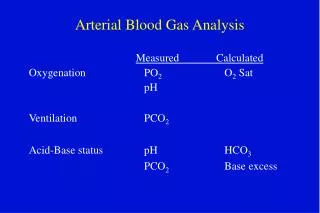
3 Processes • Ventilation (CO2) • Oxygenation (02) • Acid-Base
4 Equations 3 Physiologic Processes • EquationPhysiologic Process • 1) PaCO2 equation ------------------------------------- Alveolar ventilation • 2) Alveolar gas equation ----------------------------- Oxygenation • 3) Oxygen content equation ------------------------- Oxygenation • 4) Henderson-Hasselbalch equation-------------- Acid-base balance
PaCO2 Equation VCO2 = CO2 production VA = VE – VD VE = minute (total) ventilation VD = dead space ventilation 0.863 converts units to mm Hg VCO2 x 0.863 PaCO2 = ----------------- VA PaCO2 reflects ratio of metabolic CO2 production to alveolar ventilation
VCO2 (0.863) = PaCO2 Ventilation
PaCO2 Blood Alveolar Ventilation • >45 mm Hg ------- Hypercapnia ----- Hypoventilation • 35 - 45 mm Hg –- Eucapnia ---------- Normal ventilation • <35 mm Hg ------- Hypocapnia ------- Hyperventilation
CO2 production PaCO2 Alveolar Ventilation
Hypercapnea • The only physiologic reason for elevated PaCO2 is inadequate alveolar ventilation (VA) for the body’s CO2 production (VCO2) • Hypercapnia can arise from insufficient total ventilation, increased dead space, or a combination of the two
Case • 54 yr female presents with acute SOB • Post colon resection for a malignant bowel obstruction • Shortly after returning home from hospital she experienced sudden chest pain worse with inspiration.
Pulmonary Embolus CO2 production PaCO2 (↑ ↑ ventilation) – (↑dead space)
Alveolar Gas Equation PAO2 = PIO2 - 1.2 (PaCO2) Alveolar O2 = P Inspired O2 –Arterial CO2 • PIO2 = FIO2 (PB – 47 mm Hg) • PIO2 = Inspired O2 (Barometric Pressure – 47 mm Hg) • water vapor pressure at normal body temperature
This describes the factors that influence O2 in the alveoli • Almost always, • Alveolar O2 is higher than Arterial O2
Alveolar O2 = P Inspired O2 –Arterial CO2 • PIO2 = Inspired O2 (Barometric Pressure – 47mm Hg) • Thus, when PAO2 ↓, PaO2 ↓
Why do I care? • If everything else is constant… • as ↑PaCO2 both PAO2 and PaO2 will decrease • (hypercapnia causes hypoxemia) • as ↓FIO2 both PAO2 and PaO2 will decrease • (suffocation causes hypoxemia) • as ↓PB (e.g., with altitude) both PAO2 and PaO2 will decrease • (mountain climbing causes hypoxemia)
P(A-a)O2 the “A-a gradient” • P(A-a)O2 is the alveolar-arterial difference in pO2 • It results from gravity-related blood flow changes within the lungs (normal ventilation-perfusion imbalance) • PAO2 is always calculated • PaO2 is always measured
P(A-a)O2 the “A-a gradient” • Normal P(A-a)O2 ranges from @ 5 to 25 mm Hg ORA • (it increases with age) • A higher than normal P(A-a)O2 means the lungs are not transferring oxygen properly from alveoli into the pulmonary capillaries • An ↑P(A-a)O2 signifies a problem within the lungs • Exception: right to left cardiac shunts
NON-RESPIRATORY P(A-a)O2Cardiac right to left shunt Increased Decreased PIO2 NormalLow mixed venous oxygen content* Increased RESPIRATORYPulmonary right to left shunt IncreasedVentilation-perfusion imbalance IncreasedDiffusion barrier IncreasedHypoventilation (increased PaCO2) Normal *Unlikely to be clinically significant unless there is right to left shunting or ventilation-perfusion imbalance
Ventilation-Perfusion imbalance • A normal amount of ventilation-perfusion (V-Q) imbalance accounts for the normal P(A-a)O2 • By far the most common cause of low PaO2 is an abnormal degree of ventilation-perfusion imbalance within the millions of alveolar-capillary units • Virtually all lung disease lowers PaO2 via V-Q imbalance • i.e. asthma, pneumonia, atelectasis, pulm edema, COPD • Diffusion barrier is seldom a major cause of low PaO2 • (it can lead to a low PaO2 during exercise)
Case • 34 Male presents with HA • Working out of town – sleeping in the shop • Awoken at 2-3am by an alarm but went back to sleep • Found by his foreman at about 9am • Drove back to Calgary but had difficulty staying awake
How much oxygen is in the blood?PaO2 vs. SaO2 vs. CaO2 • OXYGEN PRESSURE: PaO2 • PaO2 reflects only free oxygen molecules dissolved in plasma and not those bound to hemoglobin • PaO2 cannot tell us “how much” oxygen is in the blood • OXYGEN SATURATION: SaO2 • The percentage of all the available heme binding sites saturated with oxygen is the hemoglobin oxygen saturation (in arterial blood, the SaO2) • How much hemoglobin is there? • OXYGEN CONTENT: CaO2 • CaO2 is the only value that incorporates the hemoglobin content (units ml O2/dl) • Oxygen content can be measured directly or calculated by the oxygen content equation: • CaO2 = (Hb x 1.34 x SaO2) + (.003 x PaO2)
Case Continued • returned a few hours later with mental confusion • this time both SaO2 and COHb were measured
CO has a ‘double-whammy’ effect • decreases SaO2 by the amount of COHb present • shifts the O2-dissociation curve to the left, retarding unloading of oxygen to the tissues • CO does not affect PaO2, only SaO2 • To detect CO poisoning, SaO2 and/or COHb must be measured • In the presence of excess CO, SaO2 will be lower than expected from the PaO2
Carbon Monoxide • CO is colorless, odorless gas, a product of combustion; all smokers have excess CO in their blood, typically 5-10% • CO binds 200x more avidly to hemoglobin than O2, effectively displacing O2 from the heme binding sites • CO is a major cause of poisoning deaths world-wide • Normal %COHb in the blood is 1-2%, from metabolism and small amount of ambient CO • (higher in smokers and traffic-congested areas)
SaO2 and CaO2: test your understanding Below are blood gas results from four pairs of patients. For each letter pair, state which patient, (1) or (2), is more hypoxemic. Units for hemoglobin content (Hb) are gm% and for PaO2 mm Hg. a) (1) Hb 150, PaO2 100, pH 7.40, COHb 20% (2) Hb 120, PaO2 100, pH 7.40, COHb 0 b) (1) Hb 150, PaO2 90, pH 7.20, COHb 5% (2) Hb 150, PaO2 50, pH 7.40, COHb 0
SaO2 and CaO2: test your understandingAnswers a) (1) CaO2 = .78 x 15 x 1.34 = 15.7 ml O2/dl (2) CaO2 = .98 x 12 x 1.34 = 15.8 ml O2/dl The oxygen contents are almost identical, and therefore neither patient is more hypoxemic. However, patient (1), with 20% CO, is more hypoxic than patient (2) because of the left-shift of the O2- dissociation curve caused by the CO b) (1) CaO2 = .87 x 15 x 1.34 = 17.5 ml O2/dl (2) CaO2 = .85 x 15 x 1.34 = 17.1 ml O2/dl A PaO2 of 90 mm Hg with pH of 7.20 gives an SaO2 of @ 92%; subtracting 5% COHb from this value gives a true SaO2 of 87%, used in the CaO2 calculation of patient (1). A PaO2 of 50 mm Hg with normal pH gives an SaO2 of 85%. Thus patient (2) is slightly more hypoxemic.
Acid-Base • Normal serum pH is between 7.36-7.44 • A pH outside 6.8 – 7.8 is incompatible with life • pH is maintained by 3 systems • Physiologic buffers • Lungs • Kidneys • Disorders in any of these systems leads to alterations in blood pH
Physiologic Buffers • 1) Bicarbonate-carbonic acid • H+ + HCO3- ↔ H2CO3 ↔ H2O + CO2 • 2) Blood protein buffers • Hemoglobin • 3) Bone • Reservoir of bicarb and phosphate
Lungs • ∆ pH sensed by peripheral and central chemoreceptors • Peripherally (carotid bodies) • Centrally (medulla oblongata) • ↓ pH • Increased minute ventilation • Lowers PaCO2 • ↑ pH • Decreased ventilatory effort • Increases PaCO2
Kidneys • Not involved in acute compensation • After 6hrs of Alkalemia • Excretion of HCO3- • Retention of H+ • 6-12hrs Acidosis • Excretion of H+ • Retention of HCO3-
Terminology • Acidemia: blood pH < 7.35 • Acidosis: a physiologic process that, occurring alone, tends to cause acidemia • e.g.: metabolic acidosis from decreased perfusion (lactic acidosis); respiratory acidosis from hypoventilation • If the patient also has an alkalosis at the same time, the resulting blood pH may be low, normal or high
Terminology • Alkalemia: blood pH > 7.45 • Alkalosis: a primary physiologic process that, occurring alone, tends to cause alkalemia • i.e.: metabolic alkalosis from excessive diuretic therapy; respiratory alkalosis from acute hyperventilation • If the patient also has an acidosis at the same time, the resulting blood pH may be high, normal or low.
Terminology • Primary acid-base disorder: One of the four acid-base disturbances that is manifested by an initial change in HCO3- or PaCO2. • Compensation: The change in HCO3- or PaCO2 that results from the primary event. Compensatory changes are not classified by the terms used for the four primary acid-base disturbances. • i.e. a patient who hyperventilates (lowers PaCO2) solely as compensation for MAc does not have a RAlk, the latter being a primary disorder that, alone, would lead to alkalemia. In simple, uncomplicated MAc the patient will never develop alkalemia.
Henderson without Hassel(balch) [ H+ ] x [ HCO3- ] [ H+ ] x [ HCO3- ] k1 x H2CO3 k2 x [ CO2 ] x [ H2O ] k2 x [ CO2 ] x [ H2O ] X X [ H+ ] [ HCO3- ]
Henderson without the Hassel(balch) [CO2] [ H+ ] [ HCO3- ]
Henderson-Hasselbach • Henderson's equation shows the relationship between [H+], [HCO3-], and PCO2 • It performs the same function as the more Henderson-Hasselbalch Equation
Acid-Base Disorders • Respiratory disorders • Alter the serum PaCO2 • Metabolic disorders • Alter the serum HCO3- Thanks Marc!
ACIDOSIS Hypoventilation Pulmonary pathology Airway obstruction Decreased respiratory drive ALKALOSIS Hyperventilation CNS disease Hypoxemia Anxiety Toxic states Hepatic insufficiency Assisted ventilation Respiratory Disorders Thanks Marc!
ACIDOSIS Wide gap metabolic acidosis Non-AG metabolic acidosis ALKALOSIS Saline responsive Saline resistant Metabolic Disorders Thanks Marc!
Addition of Acids or Creation of Acids CAT MUDPILES Carbon monoxide/cyanide Alcohol/AKA Toluene Methanol Uremia DKA Paraldehyde INH/Iron Lactic Acidosis Ethylene glycol Salicylates Anion Gap Metabolic Acidosis Thanks Marc!
Excessive loss of HCO3- OR Inability to excrete H+ HARD UPS Hyperalimentation/Hyperventilation Acids/Addison’s/Acetazolamide RTA Diarrhea/Dehydration/ Diuretics Uterosigmoidostomy Pancreatic fistula or drainage Saline (large amounts) Normal AG Metabolic Acidosis Thanks Marc!
Metabolic Alkalosis • Saline Responsive • Vomit → lose HCl • Kidneys try to hang on to H+ • …excrete Na+ instead • Until, dehydration kicks in → Renin/Aldo • If we rehydrate we allow the kidneys to work