Download
1 / 55
580 likes | 1.31k Vues
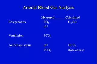
Arterial Blood Gas Analysis. October 27, 2009 Prepared by Dr. Trey Woods D.O. Presented by Dr. Nick Cardinal D.O. What is an ABG?. The Components pH / PaCO 2 / PaO 2 / HCO 3 / O 2 sat / BE Desired Ranges pH - 7.35 - 7.45 PaCO 2 - 35-45 mmHg PaO 2 - 80-100 mmHg HCO 3 - 21-27

E N D
Arterial Blood Gas Analysis October 27, 2009 Prepared by Dr. Trey Woods D.O. Presented by Dr. Nick Cardinal D.O.
What is an ABG? • The Components • pH / PaCO2 / PaO2 / HCO3 / O2sat / BE • Desired Ranges • pH - 7.35 - 7.45 • PaCO2 - 35-45 mmHg • PaO2 - 80-100 mmHg • HCO3 - 21-27 • O2sat - 95-100% • Base Excess - +/-2 mEq/L
ABG analysis • Why do we care ? • Critical care requires a good understanding • Helps in the differential and final diagnosis • Helps in determining treatment plan • Treating acid/base disorders helps medications work better (i.e. antibiotics, vasopressors, etc.) • Helps in ventilator management • Severe acid/base disorders may need dialysis • Changes in electrolyte levels in acidosis (increased K+ and Na+, and decreases in HCO3)
Logistics • When to order an arterial line -- • Need for continuous BP monitoring • Need for multiple ABGs • Where to place -- the options • Radial • Femoral • Brachial • Dorsalis Pedis • Axillary
Acid Base Balance • The body produces acids daily • 15,000 mmol CO2 • 50-100 mEq Nonvolatile acids • The lungs and kidneys attempt to maintain balance
Acid Base Balance • Assessment of status via bicarbonate-carbon dioxide buffer system • CO2 + H2O <--> H2CO3 <--> HCO3- + H+ • ph = 6.10 + log ([HCO3] / [0.03 x PCO2])
The Terms • BASES • Alkalemia • Alkalosis • Respiratory ↓CO2 • Metabolic ↑HCO3 • ACIDS • Acidemia • Acidosis • Respiratory ↑CO2 • Metabolic ↓HCO3
Transport of CO2 In the Blood • At rest 100ml of blood transports 4 ml CO2 from tissue to lungs. CO2 in blood affects acid-base balance. • CO2 is transported in blood in three forms • Dissolved CO2, bicarb, comb with proteins as carbamino cmpds. • Dissolved: 0.36ml of CO2 /100ml blood (9% of all) • Bicarbonate: reacts wit water in blood to form carbonic acid and then bicarb (accounting for 60-70% of CO2 transport to tissues). • Carbonic acid formed in RBC dissoc into H+ and bicarb ions. • H+ bind to Hb making it reduced, a better proton acceptor, this helps in peripheral blood with loading of CO2. • Deoxygenation of blood increases its ability to carry CO2: Haldance Effect
Transport of CO2 In the Blood • Carbaminohemoglobin and carbaminoproteins: Are formed from CO2 with terminal amine groups in blood proteins. Ie globin of hemoglobin HbCO2 • This occurs with a very loose bond allowing easy release in the alveoli, where the PCO2 lower than tissue capillaries. Unloading of O2 in peirpheral capillaries facilitates the loading of CO2 . • CO2 can be carried to the lungs in combo with Hb about 20-30% of total, about 1.5ml of CO2 in each 100 ml of blood.
Change in Acidity during CO2 Transport • CO2 transport has profound effect on acid-base status of blood. • Partial pressure of arterial oxygen • PaO2 normal 90-100mm Hg • PaO2 reflects the functional capabilities of the lungs and determines the rate at which O2 enters the tissues. • Factors that affect PaO2: • Va, FIO2, Altitude, age, and oxyhb dissoc. curve
Partial pressure of arterial oxygen • Alveolar Ventilation (Va) • If the pt hyperventilates PaCO2 tends to fall and PaO2 rise • If PaCO2 Falls by 1mm Hg then PaO2 rises by 1.-1.2mmHg • This is how lungs make up for some pulm dysfuntion, by hyperventilating. • FIO2 is often not considered well in evaluating PaO2 • O2 by nasal cannula actual delivered FIO2 is 25-30%. • Mask inhaled FIO2 is usually half that delivered to the mask • EXPECTED PaO2 multiply percentage by 6. • ie 60% O2 x 6=360mm HG
Partial pressure of arterial oxygen • Altitude • Greater altitude, the lower the PO2 • PaO2 drops about 3-4mm Hg for each 1000 foot rise above sea level. • Ie 30,000 feet inspired air air at barometric pressure of 226 mm Hg PaO2 is only 21mm Hg • Age • PaO2 drops 3-4mm Hg per decade after the patient reaches 20-30 yrs of age.
Alveolar-Arterial Oxygen difference • Determine degree to which lung function is impaired: use [P(A-a)O2] • Usually P(A-a)O2 should be 10 plus 1/10th pt’s age. • P(A-a)O2 of 20-30 mm Hg mild pulm dysfuntion • P(A-a)O2 of greater than 50 mm Hg on room air usually indicates severe pulmonary dysfunction. The cause of Increase A-a gradient include: • Intrapulmonary shunt (less ventilation than perfusion, or a V/Q ratio) intracardiac shunt, and diffusion abnormalities.
Shunting in the lung • Four types of alveolar capillary units • 1st vent and perfuse are normal then unit is normal • 2nd is if there is vent without perfuse, the unit is considered to be dead space (of high V/Q) • 3rd if there is perfuse without vent, unit is RL shunt or (low V/Q • 4th if there is neither vent nor perfuse, unit is silent • Shunt is the fraction of blood passing the lung without being oxygenated.
Oxygen Content • O2 is carried in blood either dissolved or on hb. • Fully saturated hb can carry 1.34ml of O2 , thus a pt with hb 15 can carry 20.1 ml of O2 per 100ml • PaO2
Oxyhemoglobin Saturation • PaO2 of 60mm Hg correlates to and SaO2 of 90% • Hb is 15 and tissue removes 5.0ml of oxygen from each 100ml of blood. • Factors that affect curve • Left shift • Alkalosis, hypothermia, abnormal and fetal hemoglobin, carboxyhemoglobin, methemoglobin, CO, Decreased 2,3 DPG • Right Shift • Acidosis, Increased Pco2 • Hyperthermia • Increased 2,3 DPG
The Anion Gap • Na – (Cl + HCO3) • NaHCO3 + HCL NaCL + H2CO3 • NaHCO3 + HX NaX+ H2CO3 • Unmeasured cations: calcium, magnesium, gamma globulins, potassium. • Unmeasured anions: albumin, phosphate, sulfate, lactate.
Gap Acidosis • Methanol • Uremia • Diabetic ketoacidosis • Paraldehyde • INH • Lactic acidosis • Ethylene Glycol • Salicylate
Non Gap Acidosis • H: hyperalimentation • A: acetazolamide • R: RTA • D: diarrhea • U: rectosigmoidostomy • P: pancreatic fistula
Metabolic Acidosis • Respiratory compensation process takes 12-24 hours to become fully active. Protons are slow to diffuse across the blood brain barrier. In the case of LA this will be faster because LA is produced in the brain. • The degree of compensation can be assessed by using Winter’s Formula. It is INAPPROPRIATE to use this formula before the acidosis has existed for 12-24 hours. • PCO2 = 1.5 (HCO3) + 8 +/-2. • ↓pH, ↓HCO3
Decreased anion gap • Decrease in unmeasured anions • Hypoalbuminemia • Increase in unmeasured cations • Hypercalcemia • Hypermagnesemia • Hyperkalemia • Multiple myeloma • Lithium toxicity
Metabolic Alkalosis • Generation by gain of HCO3 and maintained by abnormal renal HCO3 absorption. • This is almost always secondary to volume contraction (low Cl in urine, responsive to NaCl, maintained at proximal tubule) • Vomiting: net loss of H+ and gain of HCO3. • Diuretics: ECFV depletion • Chronic diarrhea: ECFV depletion • Profound hypokalemia • Renal failure: if we cannot filter HCO3 we cannot excrete it. • Mineralocorticoid excess: increased H secretion, hypokalemia (Na/K exchanger), saline resistant).
Metabolic Alkalosis • ↑pH, ↑HCO3 • ↑PCO2 by 0.7 for every 1mEq/L ↑ in HCO3
Respiratory Acidosis • Acute or Chronic: has the kidney had enough time to partially compensate? • The source of the BUFFER (we need to produce bicarb) is different in these states and thus we need to make this distinction. • ↓ph, ↑CO2, ↓Ventilation
Respiratory Acidosis • Acute : H is titrated by non HCO3 organic tissue buffers. Hb is an example. The kidney has little involvement in this phase. • 10 mm Hg increase in CO2 / pH should decrease by .08 • Chronic: The mechanism here is the renal synthesis and retention of bicarbonate. As HCO3 is added to the blood we see that [Cl] will decrease to balance charges. • This is the hypochloremia of chronic metabolic acidosis. • 10 mm H increase in CO2 / pH should decrease by .03
Respiratory Acidosis • Elevation of CO2 above normal with a drop in extracellular pH. • This is a disorder of ventilation. • Rate of CO2 elimination is lower than the production • 5 main categories: • CNS depression • Pleural disease • Lung diseases such as COPD and ARDS • Musculoskeletal disorders • Compensatory mechanism for metabolic alkalosis
Respiratory Alkalosis • Initiated by a fall in the CO2 activate processes which lower HCO3. • Associated with mild hypokalemia. Cl is retained to offset the loss of HCO3 negative charge. • Acute response is independent of renal HCO3 wasting. The chronic compensation is governed by renal HCO3 wasting. • Causes • Intracerebral hemorrhage • Drug use : salicylates and progesterone • Decreased lung compliance Anxiety • Liver cirrhosis • Sepsis
Mixed Acid-Base Disorders • Patients may have two or more acid-base disorders at one time • Delta Gap Delta HCO3 = HCO3 + Change in anion gap >24 = metabolic alkalosis
Arterial Blood Gas (ABG) Analysis • ABG interpretation Follow rules and you will always be right !! 1) determine PH acidemia or alkalemia 2) calculate the anion gap 3) determine Co2 compensation (winters formula) 4) calculate the delta gap (delta HCO3)
ABG analysis • Arterial Blood Gas (ABG) –interpretation • Always evaluate PH first • Alkalosis – PH > 7.45 • Acidosis – PH < 7.35 • Determine anion gap (AG) – AG = NA – (HCO3+ CL) • AG metabolic acidosis • Non AG acidosis – determined by delta gap • Winters formula • Calculates expected PaCO2 for metabolic acidosis • PaCO2 = 1.5 x HCO3 + 8
ABG analysis • Delta gap • Delta HCO3 = HCO3 (electrolytes) + change in AG • Delta gap < 24 = non AG acidosis • Delta gap > 24 = metabolic alkalosis • Note: The key to ABG interpretation is following the above steps in order.
Look at the pH. • pH < 7.35, acidosis • pH > 7.45, alkalosis Look at PCO2, HCO3- • Main pathology will be the change correlates with the pH. • If alkalosis pCO2 will be low or Bicarb high • If acidosis pCO2 will be high or Bicard low • The other abnormal parameter is the compensator response Respiratory or Metabolic • pCO2 - respiratory • Bicarb - metabolic
Metabolic Acidosis? Anion Gap? • >12 - ketoacidosis, uremia, lactic acidosis, or toxins • Delta ratio to check for gap and non gap disorders , or metabolic alkalosis happening simultaneously • Normal anion gap - diarrhea OR unknown. If unknown calculate urine anion gap, if positive likely RTA, if neg liekly diarrhea Metabolic Alkalosis If urin Cl is > 20 it is chloride-resistant alkalosis (increased mineralcorticoid activity If <20 chloride responsive alkalosis (vomitting or gastric loss)
Sample Problem #1 • An ill-appearing alcoholic male presents with nausea and vomiting. • ABG - 7.4 / 41 / 85 / 22 • Na- 137 / K- 3.8 / Cl- 90 / HCO3- 22
Sample Problem #1 • Anion Gap = 137 - (90 + 22) = 25 anion gap metabolic acidosis • Winters Formula = 1.5(22) + 8 ± 2 = 39 ± 2 compensated • Delta Gap = 25 - 10 = 15 15 + 22 = 37 metabolic alkalosis
Sample Problem #2 • 22 year old female presents for attempted overdose. She has taken an unknown amount of Midol containing aspirin, cinnamedrine, and caffeine. On exam she is experiencing respiratory distress.
Sample Problem #2 • ABG - 7.47 / 19 / 123 / 14 • Na- 145 / K- 3.6 / Cl- 109 / HCO3- 17 • ASA level - 38.2 mg/dL
Sample Problem #2 • Anion Gap = 145 - (109 + 17) = 19 anion gap metabolic acidosis • Winters Formula = 1.5 (17) + 8 ± 2 = 34 ± 2 uncompensated • Delta Gap = 19 - 10 = 9 9 + 17 = 26 no metabolic alkalosis
Sample Problem #3 • 47 year old male experienced crush injury at construction site. • ABG - 7.3 / 32 / 96 / 15 • Na- 135 / K-5 / Cl- 98 / HCO3- 15 / BUN- 38 / Cr- 1.7 • CK- 42, 346
Sample Problem #3 • Anion Gap = 135 - (98 + 15) = 22 anion gap metabolic acidosis • Winters Formula = 1.5 (15) + 8 ± 2 = 30 ± 2 compensated • Delta Gap = 22 - 10 = 12 12 + 15 = 27 mild metabolic alkalosis
Sample Problem #4 • 1 month old male presents with projectile emesis x 2 days. • ABG - 7.49 / 40 / 98 / 30 • Na- 140 / K- 2.9 / Cl- 92 / HCO3- 32
Sample Problem #4 • Metabolic Alkalosis, hypochloremic • Winters Formula = 1.5 (30) + 8 ± 2 = 53 ± 2 uncompensated
ABG analysis • 33 y/o with DKA presents with the following: • Na = 128, Cl = 90, HCO3 = 4, Glucose = 800 • 7.0/14/90/4/95% • PH = acidemia • AG = 128 – (90 + 4) = 34 • Winters formula – 1.5(4) + 8 = 14 • Delta gap = 4 + (34 – 12) = 26
ABG analysis • Answer • AG acidosis with appropriate respiratory compensation • History c/w ketoacidosis secondary to DKA with appropriate respiratory compensation
ABG analysis • 56 y/o with COPD exacerbation and hypotension and associated diarrhea x 7 days presents with the following ABG: • 7.22/30/65/10/90% • PH(7.22) = acidemia • AG = 139 – (10 + 110) = 19 (nl AG = 8-12) • Winters formula • PaCO2 = 1.5 (HCO3) + 8 = 1.5 (10) + 8 = 23 • Delta gap • Delta gap = HC03 + change in the AG = 24 • Delta gap = 10 + (19 – 12) = 10 + 7 = 17 • Delta gap = 17 139 110 20 120 4.0 10 1.5
ABG - example • Triple disorder • AG acidosis - • Incomplete respiratory compensation • Non AG acidosis • History would suggest AG acidosis is secondary to hypotension with lactic acid build up and the patient is not able to compensate with his COPD therefore there is no respiratory compensation and the non AG acidosis is secondary to diarrhea with associated HCO3 loss.
Example # 1 • 44 yo M 2 weeks post-op from total proctocolectomy for ulcerative colitis. • Na+ 134, K+ 2.9, Cl- 108, HCO3- 16, BUN 31, Cr 1.5 • BG: 7.31/ 33 /93 / 16