TRANSLATION
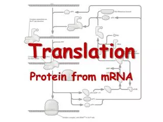
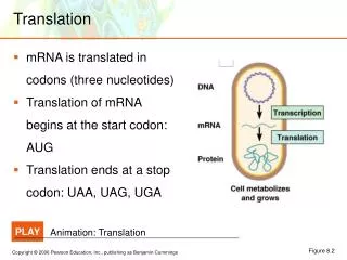
TRANSLATION. Dr.S.Chakravarty MD. Site :-Ribosomes in Cytoplasm . TRANSLATION . Dr.S.Chakravarty , MBBS,M.D . CODONS. A sequence of three nucleotides which together form a unit of genetic code in a DNA or RNA molecule . SEQUENCE OF THREE NUCLEOTIDES CODING FOR AN AMINO ACID.

TRANSLATION
E N D
Presentation Transcript
TRANSLATION Dr.S.Chakravarty MD
TRANSLATION Dr.S.Chakravarty , MBBS,M.D
CODONS A sequence of three nucleotides which together form a unit of genetic code in a DNA or RNA molecule. SEQUENCE OF THREE NUCLEOTIDES CODING FOR AN AMINO ACID
Features of Genetic code • Degeneracy – multiple codons must decode same amino acid. • 61 of the total 64 codons – code for amino acids • only 20 aminoacids. • 3 codons – stop codons (UAA, UAG, UGA).
2. Unambiguous – for any specific codon only 1 aminoacid is indicated. AUG – always codes for methionine. 3. Non-overlapping : AUGUUU – codes for methionine and phenylalanine.
4. Without punctuations – no spacing or comma between two codons. It is continuous and nonstop from 5’end to 3’end. 5. Universal : triplet codon is the same for a particular aminoacid in all the living species. Exception: mitochondrial DNA varies.
6. For those amino acids having more than one codon, the first two bases in the codon are usually same. CUU CUC CUA CUG Leucine
Wobble hyothesis • States that rules of base pairing are relaxed at the third position, so that a base can pair with more than one complementary base. • Some tRNA anticodons have Inosine at the 5’ end. Inosine can pair with U, C, or A of the 3rd nucleotide of the genetic code. At least one species of t-RNA exist for for each of the aminoacid
21st and 22nd amino acids • Selenocysteine • Pyrrolysine No genetic code available?
How does amino acids selenocysteine and pyrrolysine gets inserted into protein without genetic code ? • UGA – stop codon • SECIS – SElenoCysteineInsertion Sequence • Present in the 3’UTR region next to stop codon UGA will help the t-RNA to the sequence for selenocysteine. • UAG – stop codon • PYLIS downstream sequence in 3’UTR sequence
TACGCACATTTACGTACG DNA aa aa aa aa aa aa aa AUGCGUGUAAAUGCAUGC mRNA protein trait Mutations • Changes to DNA are called mutations • change the DNA • changes the mRNA • may change protein • may change trait
What Causes Mutations? • There are two ways in which DNA can become mutated: • Mutations can be inherited. • Parent to child • Mutations can be acquired. • Environmental damage • Mistakes when DNA is copied
Types of mutations • Changes to the letters (A,C,T,G bases) in the DNA • point mutation • change to ONE letter (base) in the DNA • may cause change to protein, may not • frameshift mutation • addition of a new letter (base) in the DNA sequence • deletion of a letter (base) in the DNA • both of these shift the DNA so it changes how the codons are read • big changes to protein!
Point Mutations • One base change • can change the meaning of the whole protein THEFATCATANDTHEREDRATRAN Does this changethe sentence? A LITTLE! THEFATCARANDTHEREDRATRAN OR THEFATCATANDTHEREDBATTAN
Single base changes may be of two types :- • Transition Purine to Purine , Pyr to Pyr • TransversionPurine to Pyrimidine or vice versa
Doesthis changethe protein? DEPENDS…
Effects of single base changes • Silent mutation :- No detectable change . • Generally here the third nucleotide of a codon • Wobble hypothesis • Missense effect :- If a different amino acid is incorporated • Acceptable • Partially acceptable • Unacceptable • Nonsense codon:- Premature termination of the peptide chain
Point Mutations • Silent mutation = no change to protein AUGCGUGUAUACGCAUGCGAGUGA MetArgValTyrAlaCysGluStop AUGCGUGUAUACGCUUGCGAGUGA MetArgValTyrAlaCysGluStop
Point Mutations • Missense mutation = changes amino acid AUGCGUGUAUACGCAUGCGAGUGA MetArgValTyrAlaCysGluStop AUGCGUGUAUACGUAUGCGAGUGA MetArgValTyrValCysGluStop
Frameshift Mutations • Add or delete one or more bases • changes the meaning of the whole protein THEFATCATANDTHEREDRATRAN THEFATCANTANDTHEREDRATRAN OR THEFATCAANDTHEREDRATRAN
Frameshift Mutations • Addition = add one or more bases AUGCGUGUAUACGCAUGCGAGUGA MetArgValTyrAlaCysGluStop AUGCGUGUAUACGUCAUGCGAGUGA MetArgValTyrValMetArgValA
Frameshift Mutations • Deletion = lose one or more bases AUGCGUGUAUACGCAUGCGAGUGA MetArgValTyrAlaCysGluStop AUGCGUGUAUACGAUGCGAGUGA MetArgValTyrAspAlaSerGA
Cystic fibrosis • Broken salt channel in cells • strikes 1 in 2500 white births • gene codes for a protein channelthat allows salt to flow across cell membrane • broken protein doesn’t work as channel • doesn’t allow salt out of cell, so water doesn’t flow out either • thicker & stickier mucus coating around cells • mucus build-ups in lungs & causes bacterial infections • destroys lung function • without treatment children die before 5; with treatment can live past their late 20s
Effect on Lungs Salt channel transports salt through protein channel out of cell Osmosis problems! normal lungs airway salt salt channel normal mucus H2O cells lining lungs cystic fibrosis salt thick mucus H2O mucus & bacteria build up= lung infections & damage
Deletion leads to Cystic fibrosis deletion Loss of one amino acid!
NEED A MUTATION FOR SUPERFAST LEARNING !!
Suppressor Mutations Suppressor t RNA molecules are formed as a result of alterations in the anti codon regions capable of suppressing certain missense mutations. They can counteract some effects of mutations. They are abnormal t RNA molecules. Nonsense suppressor t RNA can allow can suppress the normal termination signals to allow an undesirable read through
Structure of t-RNA Antiparallel to the coding sequence of amino acid on the m-RNA. Recognition of t-RNA to the enzyme aminoacyl t-RNA synthatase Binding of aminoacyl-tRNA to ribosomal surface at the site of protein synthesis. Site of attachment of specific amino acid Sequence of code in anticodon arm : 3’-variable base-modified purine-XYZ-pyr, pyr-5’
Activation of t-RNA Step 1: amino acid + ATP → aminoacyl-AMP + Ppi Step 2: aminoacyl-AMP + tRNA → aminoacyl-tRNA + AMP Twenty different aminoacyl t-RNA synthases for twenty different aminoacids
Ribosomes • consist of ribosomal proteins and ribosomal RNAs (rRNAs). • have a large subunit and a small subunit. • The rRNAs provide for important catalytic functions associated with translation.
tRNAs • 75–90 nucleotides long and contain posttranscriptionally modifiedbases. • 2-dimensional structure of tRNAs is a cloverleaf.
Activation of t-RNA Step 1: amino acid + ATP → aminoacyl-AMP + Ppi Step 2: aminoacyl-AMP + tRNA → aminoacyl-tRNA + AMP Twenty different aminoacyl t-RNA synthases for twenty different aminoacids
Translation of mRNA Can Be Divided into Three Steps • Initiation • Elongation • Termination
Initiation requires: • the small and large ribosomal subunits • mRNA • GTP • charged initiator tRNA • initiation factors
Protein synthesis – Eukaryotes • Initiation • Elongation • Termination • m-RNA is read from 5’ 3’ end. • m-RNA is identified by 18 s RNA through 7 methyl guanosine CAP
Initiation • Formation of 43 s pre-initiation complex: a)First the ribosome dissociates into into 40S and 60S subunits. 40 S subunit 5 3 1A Initiation factors eIF3 and eIF1A and eIF5 bind to the dissociated 40 S unit GTP 2 Binary complex met tRNAi 40 S subunit 5 43 s pre-initiation complex 3 1A 2
Role of eIF-2 α • eIF-2α is regulated by kinases. • Phosphorylation by at least four different protein kinases during stress: • Starvation • Viral infection • Abnormal protein (misfolded proteins) • Hyperosmolarity • Heat shock • Phosphorylated eIF-2α binds tightly to and inhibits eIF-2β which is required for regeneration of GTP from GDP on factor 2 • Insulin increases protein sysnthesis by removing the phosphate and inactivating eIF-2a
2. Formation of 48s initiation complex 40 S subunit 5 CAP (A)n 3 4F =4E+ 4G4A ATP 1A 2 4F CAP (A)n 4B ATP Cap helps in binding m- RNA to 43S preinitiation complex 4F CAP (A)n M-RNA 4B 4F (A)n CAP 5 3 1A 2 48s initiation complex
Kozak Consensus Sequence is a sequence which occurs on eukaryoticmRNA and has the consensus (gcc)gccRccAUGG. The Kozak consensus sequence plays a major role in the initiation of the translation process
Formation of 80s initiation complex 48s initiation complex 4F 2 Binary complex CAP 5 (A)n 3 GTP 1A 2 2 2B 60 s unit GDP eIF 5 2 (-) AUG CAP (A)n 2α A-site p Met Kozak consensus sequences P-site
Regulation of eIF – 4F • eIF 4E – helps in binding to CAP of m-RNA. It is activated by insulin which phosphorylateseIF 4E. • eIF 4G – CAP independent initiation of translation with the help of eIF 3 – which can be controlled by viruses.