Skill 304
Urine Analysis. Skill 304. Dr. Mohammad Marie Lecture 7. Learning objectives. To recognize urine analysis procedures: Physical Chemical Microscopic List some of urine crystals List some of urine casts. Results interpreting. Urine analysis. 1. 2. 3. URINE ANALYSIS .

Skill 304
E N D
Presentation Transcript
Urine Analysis Skill 304 Dr. Mohammad Marie Lecture \ 7
Learning objectives • To recognize urine analysis procedures: • Physical • Chemical • Microscopic • List some of urine crystals • List some of urine casts.
Results interpreting Urineanalysis
1 2 3
URINE ANALYSIS 1- Physical examination 2- Chemical examination 3- Microscopic examination
1- Physical examination • Color • Appearance • Urine volume • Specific gravity (SG)
NORMAL COLORE • Including color and clarity • Color : normally , pale to dark yellow
ABNORMAL COLORE Abnormal color : some drugs cause color changes 1. reddish urine: hematuria, hemoglobinuria 2. yellow-brown or green-brown urine: bilirubin cause : obstructive jaundice 3- greenish urine : infection
Appearance • Clarity: normally, clear Abnormal appearance:
Urine volume • The average adult : 1000ml to 2000ml/24h • Increased POLYURIA---more than 2000ml of urine in 24 hours 1. physiological states: water intake, some drugs, intravenous solutions 2. pathologic states: diabetes mellitus, diabetes insipidus
Urine volume • Decreased OLIGURIA : less than 400ml of urine in 24 hours ANURIA : less than 100ml of urine in 24 hours 1. pre-renal: hemorrhage, dehydration, congestive heart failure 2. post-renal: obstruction of the urinary tract (may be stones, carcinoma)
Specific gravity (SG) • Reflect the density of the urine • Range of 1.001 to 1.040 • Increased: Dehydration, Fever, Vomiting , Diarrhea , Diabetes Mellitus (urine volume↓ and SG↑) • Decreased: diabetes insipidus (urine volume↑ and SG ↓) Low fixed gravity (1.010): Renal failure
2-Chemical examination • Urine PH • Protein • Glucose • Ketones • Occult blood • Bilirubin • Urobilinogen
Urine PH • Normal PH The average is about 6 Range from 5~7 (depends on diet) • Higher PH---alkaline urine ( 7.8 – 8 .0 ) 1.drugs: sodium bicarbonate 2.vegetarian 3.alkalosis (metabolic or respiratory) 4. Infection of urinary tract • Lower PH---acid urine ( 4.5 – 5.5 ) 1. drugs: ammonium chloride 2. diabetes 3. acidosis (metabolic or respiratory)
Protein in urine • Reference value Qualitative method: negative Quantitative method: less than 150mg of protein in 24 hours • Urine proteins come from plasma protein and Tamm-Horsfall (T-H) glycoprotein
Protein in urine • Proteinuria---more than 150mg proteins in urine in 24 hours or qualitative test is positive • Proteinuria quantification (depend on the amount of protein ) heavy proteinuria---->4.0g/24 hours moderate proteinuria----1.0~4.0g/24 hours minimal proteinuria----<1.0g/24 hours
Protein in urine • Qualitative categories of proteinuria 1- Glomerularproteinuria 2- Tubular proteinuria
Glucose in urine • Reference value Qualitative method: negative • Glycosuria--- qualitative test is positive 1.hyperglycemia: diabetes mellitus Cushing’s syndrome 2.without hyperglycemia: renal tubular dysfunction, such as pyelonephritis
Ketones in urine • Including three ketone bodies: acetoacetic acid 20% acetone 2% β-hydroxybutyric acid 78% • The products of fat metabolism • Reference value: qualitative method: negative • Ketonuria--- qualitative test is positive
Ketones in urine • Ketonuria 1. diabetic ketonuria 2. nondiabeticketonuria: Hyperemesis of pregnancy Patients accompanied by vomiting or diarrhea
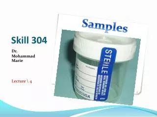
3-Microscopic examination • Sample preparation 1-Obtain fresh urine sample 2- shake the container to mix the sample 3- pipette suitable amount to test tube 4- Centrifuge it at 1500 to 3000 rpm for 5 minutes 5- Decant supernatant part 6- from the sediment Place 1 drop of urine on slide and apply cover slip 7- examine it under microscope ( 10x , 40x )
Microscopic examination • Examination A- Urine Cells B- Bacteria C- Urine Crystals D- Urine Casts
Microscopic examination A- Urine Cells : 1- Urine White Blood Cells (pus cell) Normal <2/ HPF in men and <5/ HPF in women • Few : up to 10/ HPF • Moderate : 11-40 / HPF • Many : > 40 / HPF
Microscopic examination 2- Urine Red Blood Cells : smaller and more refractile than white cells Normal <3/ HPF • Dysmorphic RBCs suggest glomerular disease
Microscopic examination 3- Epithelial cells : 3 types • Transitional epithelial cells are normally present
Microscopic examination • Squamous epithelial cells suggest contamination
Microscopic examination • Renal tubule epithelial cells suggest renal disease
Microscopic examination B- Bacteria : Diagnostic for Urinary Tract Infection • Men: Any bacteria • Women: 5 or more bacteria per HPF
Microscopic examination C-Urine Crystals 1- Calcium oxalate crystals (square envelope shape)
Microscopic examination 2- Triple phosphate crystals (coffin lid shape) • Associated with increased Urine pH (alkaline) • Associated with Proteus Urinary Tract Infection
Microscopic examination 3- Uric Acid crystals (diamond shape)
Microscopic examination D- Urine Casts 1- Epithelial cell casts of renal tubule
Microscopic examination 2- Red Blood Cell casts
Microscopic examination 3- White Blood Cell casts
Microscopic examination 4- Hyaline or mucoprotein casts
Microscopic examination 5- Granular casts
Microscopic examination 6- Waxy casts
Microscopic examination 7- Fatty casts