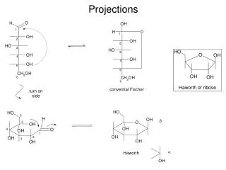
Projections
Projections. More Reactions of Sugars. Reactions of OH group(s): Esterification: Ethers:. b) Ethers (con’t). Acetals. c) Acetals (con’t). These reactions are used for selective protection of one alcohol & activation of another (protecting group chemistry).

Projections
E N D
Presentation Transcript
More Reactions of Sugars • Reactions of OH group(s): • Esterification: • Ethers:
b) Ethers (con’t) • Acetals
These reactions are used for selective protection of one alcohol & activation of another (protecting group chemistry) 1° alcohol is most reactive protect first AZT
e.g, synthesis of sucrose (Lemieux, Alberta) • Can only couple one way—if we don’t protect, get all different coupling patterns • Yet nature does this all of the time: enzymes hold molecules together in the correct orientation, BUT the mechanism still goes through an oxonium ion (more on this later)
Selectivity of Anomer Formation in Glycosides • Oxonium ion can often be attacked from both Re & Si faces to give a mixture of anomers. • How do we control this?
This reaction provides a clue to how an enzyme might stabilize an oxonium ion (see later)
Examples of Naturally Occurring di- & oligo- Saccharides Maltose: 2 units of glucose a β sugar α glycoside 1,4-linkage Lactose (milk): galactose + glucose a β sugar β glycoside 1,4-linkage
Sucrose (sugar): glucose + fructofuranose a β sugar α glycoside 1,2-glycosidic bond α-1,6-glycosidic bond Amylopectin (blood cells): an oligosaccharide α-1,4-glycosidic bond
Structure Determination of Sugars • The following is an example to review & expand your knowledge of NMR • Consider the question of glycoside formation: See NMR spectra of both anomers: They are different-diastereomers have different spectra, but which is which?
These spectra are rich in independent information: • Chemical shift, : • Reveals functional groups (see chart) • Depends on inductive effects, # of EWGs & bonds Inductive effects e.g. # of EWGs e.g. glucose– anomeric H is most downfield since 2 O atoms attached to C have more of an effect that 1 atom. we can assign the anomeric proton in the both spectra of α and β methyl glucoside bonds –alkenes, aromatics, C=O, etc
Integrals • NMR is quantitative e.g. glycosides—area under anomeric signal = 1; area under the signal at 3.3 = 3x bigger, 3 protons, must be a CH3 group • Multiplicity • Protons communicate their spins over 1, 2 or 3 bonds—reveals # of neighbors e.g. CH3-O group: a singlet, one line, no neighbors—nearst neighbor is 4 bonds away e.g. anomeric proton: a doublet, 2 peaks, one neighbor that is 3 bonds away (recall n neighbors, n +1 peaks) • Coupling Constant • Distance between peaks in a multiplet is J, coupling constant—depends on geometry
(con’t) • e.g. α glycoside: 2 peaks are 4.650-4.632 = 0.018 ppm apart spectrometer frequency = 200 MHz J = 0.018 ppm x 200 MHz = 3.6 Hz For the β-glycoside, J = 8.0 Hz • Different J values reflect different geometries: H1 – C1 – C2 – H2 = 60° in α, = 180° in β J depends on geometry according to Karplus curve: At 60°, J is small 180°, J is large J reveals the geometry, i.e., the stereochemistry of the glycoside dihedral H-X-X-H
But, looking at the spectra, note that the CHOH protons at C-2, C-3, C-4, C-5 & C-6 are all overlapping. • hard to measure each J value—How to use NMR to get a complete structure?
What about a very complex case, i.e., sucralose, the sweetener in splenda: • Where are the chlorines? Which anomer is formed? Pyranose &/or furanose ring? A challenging structure—need advanced NMR methods
Quick review of NMR theory & Pulse NMR: Not an important part of exams, but may help on questions for assignments
Modern NMR spectrometers use pulse NMR, rather than CW; advantages are: • Can acquire full spectrum in 2-3 seconds, rather than 2-3 min • Can add together data from many pulses—improves signal/noise • Can combine 2 or more pulses—allows magnetic billiards— e.g. make different CH, CH2, CH3 groups have different phase e.g. 2D NMR –COSY- to determine which H’s are coupled to one another e.g. 3D NMR –to determine protein structures & conformation in solution How does it work? We’ll do a simple treatment
Energy gap between states: More spin states in low energy (Boltzmann) = net absorption = resonance (signal!)
2-3 sec Mechanism of Absorption:
Points about FID • A sin wave with frequency -o (the difference in the frequency of RF signal & frequency emitted from nucleus) chemical shift • Decays with time as relaxation occurs (i.e., nuclei lose excitation) Watch the FID on the 200 MHz when you get your spectrum! Transform FID to Frequency domain -o (i.e. )
In a real spectrum, the FID is a complex mixture of different sine waves with • Different frequencies -o, ie., different • Different intensities, i.e. integral • Different relaxation rates, ie, different widths • FT resolves it all—based on a mathematical formula by Fourier (French mathematician 18th C) • FIDs can be added together to improve S/N: this is essential for e.g. 13C NMR FID 1 Add together: S/N improves by 2 FID 2 In general, S/N improves by 2 for each 22 = 4 times the number of scans
Going back to the spectrum of xxx: we have a problem—which CH is which & what pairs are coupled together? • Use COSY, a 2D technique that plots vs on x X y axes • Very useful—can work way around rings & assign protons A B Cross-peaks show coupled pairs Diagonal peaks B A
COSY: How does it Work? • Collect a series of spectra with different delay times: • Series of FIDs & spectra are collected and a 2D contour map is generated (contour plot)
A B Cross-peaks show coupled pairs Diagonal peaks B A • Contour plot: • H vs H • like a map • if two 1H aren’t coupled = no crosspeak
COSY tell us which protons are coupled together! Back to Sucralose • Assign anomeric H in the pyranose • Look for cross-peak → H-2 • Look for cross-peak from H-2 → H-3 etc • Extremely useful in determining chemical shift assignment • Once each H in sucralose is assigned, you can measure the coupling constants, J e.g. H-1, d, J = 4 Hz =5.37 1 or both of the H1 & H2 are equatorial H-2 3.83 (by COSY), dd, 4 & 10 Hz J2,3 = 10 Hz
H2 & H3 are both axial • H1 is equatorial: • Try 6-membered ring • Note the 2 diastereotopic protons at H6—see the coupling in the COSY spectrum • The chemical shifts are very close since they exhibit strong coupling • This is common with diastereotopic protons • Also see example in the benzoin lab (exp 7)
Other 2D Experiments • TOCSY • Correlates all the spins in a coupled spectrum, e.g. sucralose • 2 spin sets: the pyranose & the furanose rings • NOESY • Nuclear overhauser effect • Correlates protons that are close in space
NOESY is a 2D version—useful for protein conformations 13C NMR: • Usually acquired with protons decoupled • Simplifies spectra: each C 1 signal • Increased sensitivity: big nOe 1H 13C & singlet for each C (no coupling) • But lose info use DEPT • Singlet, but with attached proton info • Even better: Combine 1H & 13C (HSQC/HMBC) • Cross-peaks show which H is attached to which C (HSQC) or adjacent H’s (HMBC) • Very useful in structure determination