Download
1 / 36
380 likes | 656 Vues
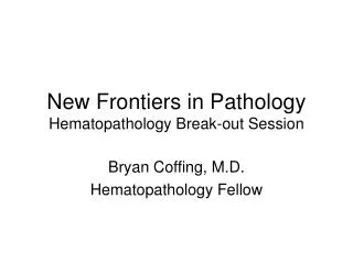
New Frontiers in Pathology. Case #5 August 3, 2012 Michael H. Roh, MD, PhD University of Michigan Health System. Disclosure Information. The speaker has no relationship representing a possible conflict of interest with respect to the content of this presentation. History.

E N D
New Frontiers in Pathology Case #5 August 3, 2012 Michael H. Roh, MD, PhD University of Michigan Health System
Disclosure Information The speaker has no relationship representing a possible conflict of interest with respect to the content of this presentation.
History • The patient is a 53 year old male with no significant medical history who presented with worsening low back pain associated with bilateral lower extremity weakness and sensory deficits. • A CT scan revealed enlarged retroperitoneal lymph nodes and an ill-defined lesion in the L5 vertebra. A CT guided FNA of the L5 lesion was performed.
Differential Diagnosis • Metastatic carcinoma • Metastatic melanoma • Metastatic germ cell tumor • Lymphoma (e.g., large cell lymphoma and Hodgkin’s lymphoma)
OCT4 PLAP c-kit D2-40
Negative Immunostains • CK7, CK20, Cytokeratin cocktail (Cam5.2/AE1/AE3) • S100, HMB45, Melan A • CD3, CD20, CD45/LCA • CD15, CD30 • AFP
Diagnosis L5 vertebral lesion, Fine-needle aspiration: • Positive for malignant cells, consistent with metastatic seminoma.
Clinical follow-up • Testicular ultrasonography: Performed 6 weeks following the FNA diagnosis. This revealed no evidence of a mass lesion in either testis. • Laboratory Tests: Serum AFP and hCG levels were not elevated. • Clinical conclusion: Metastatic seminoma vs. primary extragonadal seminoma.
Clinical follow-up • Treatment: Radiation to lumbar spine for immediate control of symptoms. Chemotherapy with 4 cycles of etoposide and cisplatin. • Follow-up: No evidence of disease progression, recurrence, and metastasis since completion of chemotherapy 2 years ago.
Extragonadal Germ Cell Tumors Stang et al. International Journal of Andrology (2012)
Extragonadal Germ Cell Tumors Variety in anatomic location (usually midline): • Brain • Pineal gland • Pituitary gland • Thymus • Mediastinum • Retroperitoneum • Pelvis • Placenta • Uterus
Incidence Rates of Retroperitoneal Germ Cell Tumors Incidence (cases per 1 million) Age (years) Stang et al. International Journal of Andrology (2012)
Hypotheses on Origin of Extragonadal Germ Cell Tumors Hypothesis #1: • Primordial germ cells originate from the proximal epiblast and migrate along the midline of the body through the hindgut to the genital ridge. • Disturbed migration of these primordial germ cells results in misplacements at different sites in the body’s midline. • These residual primordial germ cells can undergo malignant transformation.
Hypotheses on Origin of Extragonadal Germ Cell Tumors Hypothesis #2: • Extragonadal germ cell tumors represent metastases and the primary tumor regressed (“burned out”). • Histologic studies of testes of patients with extragonadal germ cell tumors reveal areas of testicular fibrosis and microlithiasis in some instances. These could represent “burned out” primary germ cell tumors.
Extragonadal Germ Cell Tumors • The histology of extragonadal germ cell tumors recapitulates that of gonadal germ cell tumors. • Seminoma (most common) • Embryonal carcinoma • Yolk sac tumor • Teratoma • Choriocarcinoma • Mixed variants
Cytology of Seminoma • Dispersed to loosely cohesive cell population. • Large tumor cells with fragile, vacuolated cytoplasm. • Round nuclei with prominent nucleoli. • Lymphocytes and granulomas in the background can be helpful clues. • Tigroid background seen in <50% of extragonadal seminomas.
Cytology of Seminoma Gupta et al. Cancer Cytopathology (2008)
Cytology of Yolk Sac Tumor • Single cells and cohesive clusters in papillary and/or acinar configurations. • Vacuolated cytoplasm. • Prominent nucleoli. • Basement membrane-like material and hyaline globules are helpful features.
Cytology of Yolk Sac Tumor Gupta et al. Cancer Cytopathology (2008)
Cytology of Embryonal Carcinoma • Cohesive clusters of large tumor cells with hyperchromatic, pleomorphic nuclei. • Scant cytoplasm. • Prominent nucleoli. • Cytologic features are less specific and difficulties can be encountered in distinguishing between embryonal carcinoma and other germ cell tumors.
Cytology of Embryonal Carcinoma Gupta et al. Cancer Cytopathology (2008)
Immunoprofiles of Germ Cell Tumors Hammerich et al. Arch Pathol Lab Med (2008)
Immunoprofiles of Germ Cell Tumors Hammerich et al. Arch Pathol Lab Med (2008)
Immunoprofiles of Germ Cell Tumors Hammerich et al. Arch Pathol Lab Med (2008)
Immunoprofiles of Germ Cell Tumors Hammerich et al. Arch Pathol Lab Med (2008)
Immunoprofiles of Germ Cell Tumors Santagata et al. Am J Surg Pathol (2008)
Immunoprofiles of Germ Cell Tumors Santagata et al. Am J Surg Pathol (2008)
Immunoprofiles of Germ Cell Tumors Santagata et al. Am J Surg Pathol (2008)
Immunoprofiles of Germ Cell Tumors Santagata et al. Am J Surg Pathol (2008)
Concluding Remarks • Germ cell tumors should be entertained in the differential diagnosis of FNAs of malignant tumors, especially in retroperitoneal and mediastinal sites. • Seminomas represent the most common extragonadal germ cell tumor and can be reliably diagnosed on FNA based on analysis of cytomorphology and immunophenotype.