Download

1 / 25
290 likes | 1.34k Vues
Cholesteatoma and chronic suppurative otitis media. Normally middle ear cleft linings: Ciliated columnar in anterior and inferior part. Cuboidal in middle part. There is nowhere lined by keratinising squamous epithelium in middle ear cleft.

E N D
Normally middle ear cleft linings: Ciliated columnar in anterior and inferior part. Cuboidal in middle part. There is nowhere lined by keratinisingsquamous epithelium in middle ear cleft. Presence of this epithelium in middle ear is called cholesteatoma(skin in the wrong place) CHOLESTEATOMA
1)Presence of congenital cell rests. 2)Invagination TM from attic or posterosuperior part of pars tensa in the form of retraction pockets(wittmacck’s theory). 3)Basal cell hyperplasia(Rueid’s theory) 4)Epithelial invasion(Habermann’s theory) 5)Metaplasia(Sade’s theory) Origin of cholesteatoma
CLASSIFICATION OF CHOLESTEATOMA 1.CONGENITAL 2.ACQUIRED,PRIMARY 3.ACQUIRED,SECONDARY
EUSTACHIAN TUBE OBSTRUCTION PERSISTENT NEGATIVE PRESSURE IN MIDDLE EAR ATTIC OR POSTERIOSUPERIOR RETRACTION POCKET PROLIFERATION OF BASAL LAYER METAPLASIA OF MIDDLE EAR MUCOSA PRIMARY ACQUIRED CHOLESTEATOMA SUBCLINICAL INFECTIONS OF MIDDLE EAR
ACUTE NECROTIZING OTITIS MEDIA REPEATED INFECTION THROUGH PERFORATION LARGE CENTRAL OR MARGINAL PERFORATION METAPLASIA OF MIDDLE EAR MUCOSA EPITHELIAL MIGRATION THROUGH PERFORATION SECONDARY ACQUIRED CHOLESTEATOMA
Expansion of cholesteatoma and destruction of bone Once cholestetoma enters middle ear cleft , it invades surrounding structures,first by following the path of least resistance, and then by enzymatic bone destruction which is caused by collagenase , acid phosphatase and proteolytic enzymes which are liberated by osteoclasts and mononuclear inflammatory cells.
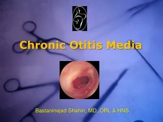
It is long standing infection of a part or whole of the middle ear cleft , characterised by ear discharge and permanent perforation . Epidemiology: Higher incidence in developing countries. Both sexes and all age group s are affected. Singlemost imp. Cause of hearing loss. Chronic suppurative otitis media
A)Tubotympanic :Also called safe or benign type . Involves antero inferior part with central perforation. B)Atticoantral: Also called unsafe or dangerous type . Involves the posterosuperior part and is associated with an attic or marginal perforation . Often associated with bone eroding process. Types of CSOM
Pathology: 1)Perforation of pars tensa 2)Middle ear mucosa 3)Polyp 4)Ossicular chain 5)Tympanosclerosis 6)Fibrosis and adhesions A)Tubotympanic type
Pseudomonas aeruginosa , proteus , E.coli and staph aerus. Bacteroidesfragilis and streptococci. Bacteriology
Clinical Features: 1)Ear discharge : non offensive mucoid or mucopurulent. 2)Hearing loss 3)Perforation 4)Middle ear mucosa:Redoedematous and swollen.
1)Examination under microscope 2)Audiogram 3)Culture and sensitivity of ear discharge 4)Mastoid X-rays. Investigations:
Aural Toilet. Ear drops. Systemic antibiotics. Precautions. Treatment of contributory causes. Surgical treatment. Reconstructive surgery. Treatment:
Pathology: Cholesteatoma Osteitis and granulation tissue Ossicular necrosis Cholesterol granuloma B)Atticoantral Type
SYMPTOMS: Ear discharge. Hearing loss. Bleeding. SIGNS: Perforation. Retraction pocket. Cholesteatoma
1)Tunning fork test 2)Audiogram 3)X-ray mastoids and CT scan of temporal bone. 4)Culture and sensitivity of ear discharge. Investigations:
1)Surgical: 2)Reconstructive surgery: 3)Conservative Treatment: Treatment:
Tubercular otitis media SYPHILITIC OTITIS MEDIA