Behandling av immunsykdommer
610 likes | 1.13k Vues
Behandling av immunsykdommer. SLE Sjøgrens syndrom Revmatoid artritt Andre autoimmune sykdommer Transplantasjonsreaksjoner. Behandling av immunsykdommer. Glucocortikoider Cytostatika Kalsinevrinhemmere mTOR hemmere Immunoglobuliner Cytokinantagonister. KROPPENS FORSVARSMEKANISMER.

Behandling av immunsykdommer
E N D
Presentation Transcript
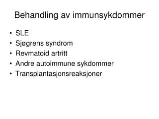
Behandling av immunsykdommer • SLE • Sjøgrens syndrom • Revmatoid artritt • Andre autoimmune sykdommer • Transplantasjonsreaksjoner
Behandling av immunsykdommer • Glucocortikoider • Cytostatika • Kalsinevrinhemmere • mTOR hemmere • Immunoglobuliner • Cytokinantagonister
KROPPENS FORSVARSMEKANISMER • Immunitet = “fri for”, “uberørt av” • Immunologi • Immunsystemet • ”Uspesifikke” forsvarsmekanismer • Ytre- og indre forsvarslinje • ”Spesifikke” forsvarsmekanismer • Cellulære forsvar (T-celler) • Antistoff-avhengige forsvar (B-celler)
Forsvare seg mot hva? • Fremmede mikrober (infeksjoner) • Bakterier • Sopp • Parasitter • Virus (eks. HIV) • Kreftceller • Utslitte/døde celler • Fremmede stoffer (eks. allergener) • Fremmed vev (transplantasjoner) • Kroppen selv: Autoimmune sykdommer! • MS, RA, o.a.
KROPPENS FORSVARSCELLER • Leukocytter • Granulocytter • Nøytrofile • Eosinofile • Basofile • Monocytter • Lymfocytter • Vevsbaserte celler • Makrofager • Makrofag-lignende celler (eks. mikroglia) • Mastceller
Uspesifikt forsvar • Naturlige/non-adaptive immunrx. • Ytre forsvarslinje • Hud, slim, nesehår, hoste/nysing • Hindrer det meste... • Magesaft, skjeden (sur pH) • Dreper de fleste mikroorganismene... • Andre mikroorganismer • Balansen i floraen av mikroorganismer • “Ufarlige” hindrer vekst av “farlige”
Uspesifikt forsvar • Indre forsvarslinje • Fagocytter (fagocytose) • Nøytrofile, makrofager • Komplementsystemet • Plasmaproteiner (MAC, opsonin) • Interferon (, , ) • Cytokin • Fra virus-infiserte celler • Beskytter ikke-infiserte celler • Akuttfaseprotein
Nøytrofile - hva “kan” de? • Amøboide bevegelser, diapedese • Kjemotakse • Fagocytose • Mediatorfrisetting • Selektiv destruksjon • Opsonin • ”Puss-celler”
Nøytrofile granula • Spesifikke granula • Små, mange, variabel fasong • Antibakterielle proteiner(fagocytiner) • Alkalisk fosfatase • Lysosomer (primære granula) • Store, runde • Hydrolytiske enzymer • Potente antibakterielle enzymer • Lysozym • Myeoloperoksydase • D-amino-oksidase
Monocytter/Makrofager • Monocytter: i blod • Makrofager: i vev • Fagocytose • Utskiller toksiske agens • O2- • Utskiller cytokiner mm. • Inflammasjon., akt. og diff. av TH • Akutt fase respons • Prosesserer og presenterer Ag til TH
Inflammatorisk respons • Makroanatomisk • rubor (rødme) • calor (varme) • dolor (smerte) • tumor (hevelse) • functio laesa (nedsatt funksjon) • Provokasjoner • Mikroorganismer, forbrenning, toksiske stoffer, fysisk overbelastning etc.
Betennelse (inflammasjon) • Første forsvarslinje • Stasjonære makrofager • Utvidelse av blodkar • Blod, fagocytter, plasmaproteiner, mediatorer, ødem, rødme, varme • Andre forsvarslinje • Innvandring av nøytrofile (kjemotakse) • Tredje forsvarslinje • Økt monocytter/makrofager (fagocytose)
Komponenter i inflammatatorisk respons • Vasodilatasjon • Økt karpermeabilitet • Kjemotaxis • Fagocytose/celledrap • Vevsreparasjon
Inflammatoriske mediatorer • Histamin • 5-HT • PAF • Bradykinin • Prostaglandiner (PGE2 o.a.) • Leukotriener (LTB4 o.a.) • NO • Cytokiner • Komplementfaktorer (C3a, C5a) • O.a.
Cytokiner • Produseres av celler i immunsystemet, endotelceller, fibroblaster o.a. • >100 løselige intercellulære signalmolekyler • Funksjoner • Immunomodulering • Proinflammatoriske, eks. interleukin (IL)-1, IL-6, TNF • Antiinflammatoriske, eks. (IL)-4, IL-10, IL-13, IFN • Differensiering, eks. IL-2, IL-4, IL-12 • Vekst, eks. IL-2, EGF • Kjemokiner, eks. IL-8, RANTES
Spesifikt forsvar • Immunreaksjon • Lymfocytter • Antigen (Ag) • Cellulære forsvar • T-lymfocytter, MHCI/II, Ag-presentering • Antistoff (Ab)-avh. forsvar (humoral) • B-lymfocytter plasmaceller Ab-prod. • Kloner, klonseleksjon, klonekspansjon • B- og Tc- hukommelsesceller
There are two classes of MHC molecules: MHC I presents cytosolic antigens (on all cells) MHC II presents endocytosed antigens (antigenpresenting cells)
The peptide fragment from the degraded antigen is loaded into a antigen-binding groove on the surface of the MHC molecule
Cytotoxic T-cells recognize infected cells (via T-cell receptor) and kill these cells by release of perforins that form channels in the target cell membrane
Cytotoxic T-cells only kill virus-infected cells with the same MHC class (MHC restriction)
CD4 and CD8 are surface proteins that acts like co- receptors for MHC I and MHC II, respectively Intracellularly, CD4 and CD8 are coupled to tyrosine kinases that participates in the activation of the T-cell
Both the B-cell antigen receptor and TCR are coupled to intra- cellular signalling pathways
When helper T-cells are stimulated via their T-cell receptor they secrete interleukins (IL2) that induce their proliferation
B-celle aktivering Plasmacelle
MHC-II APC IL-1 TNF IL-2 TH celler Plasmaceller