Download

1 / 50
530 likes | 1.22k Vues
7–1 Life Is Cellular A. The Discovery of the Cell 1. Early Microscopes 2. The Cell Theory B. Exploring the Cell C. Prokaryotes and Eukaryotes 1. Prokaryotes Eukaryotes 7–2 Eukaryotic Cell Structure A. Comparing the Cell to a Factory B. Nucleus C. Ribosomes

E N D
7–1 Life Is Cellular A. The Discovery of the Cell 1. Early Microscopes 2. The Cell Theory B. Exploring the Cell C. Prokaryotes and Eukaryotes 1. Prokaryotes Eukaryotes 7–2 Eukaryotic Cell Structure A. Comparing the Cell to a Factory B. Nucleus C. Ribosomes D. Endoplasmic Reticulum E. Golgi Apparatus F. Lysosomes G. Vacuoles H. Mitochondria and Chloroplasts 1. Mitochondria 2. Chloroplasts 3. Organelle DNA I. Cytoskeleton 7–3 Cell Boundaries A. Cell Membrane B. Cell Walls C. Diffusion Through Cell Boundaries 1. Measuring Concentration 2. Diffusion D. Osmosis 1. How Osmosis Works 2. Osmotic Pressure E. Facilitated Diffusion F. Active Transport 1. Molecular Transport 2. Endocytosis and Exocytosis 7–4 The Diversity of Cellular Life A. Unicellular Organisms B. Multicellular Organisms 1. Specialized Animal Cells 2. Specialized Plant Cells C. Levels of Organization 1. Tissues 2. Organs 3. Organ Systems Biology 1- Chapter 7 NotesPrentice Hall (pg. 168-193)
7.1: The History of the Cell Theory • Before microscopes were invented, people believed that diseases were caused by curses and supernatural spirits. • As scientists began using microscopes, they quickly realized they were entering a new world–one of microorganisms. • Microscopes enabled scientists to view and study cells, the basic units of living organisms.
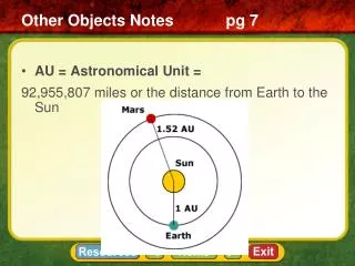
The first person to record looking at water under a microscope was Anton vanLeeuwenhoek. The microscope van Leeuwenhoek used is considered a simple light microscope because it contained one lens and used natural light to view objects. Compound light microscopes use a series of lenses to magnify objects in steps. These microscopes can magnify objects up to 1500 times. Development of Light Microscopes
The Cell Theory • Robert Hooke was an English scientist who lived at the same time as van Leeuwenhoek. • Hooke used a compound light microscope to study cork, the dead cells of oak bark. • He thought the structures he observed resembled the one room “cells” • Hooke is credited with giving cells their name • Cells are the basic building blocks of all living things.
The cell theoryis made up of three main ideas: 1. All organisms are composed of one or more cells. 2. The cell is the basic unit of structure and function of organisms. 3. All cells come from preexisting cells. Three Scientists contributed to the Cell Theory: • 1838- Schleiden: plants are made of cells • 1839- Schwann: animals are made of cells • 1855- Virchow: New cells are produced from the division of old cells
The electron microscope was invented in the 1940s. This microscope uses a beam of electrons to magnify structures up to 500,000 times their actual size. There are two basic types of electron microscopes. The scanning electron microscope scans the surface of cells to learn their three dimensional shape. The transmission electron microscope allows scientists to study the structures contained within a cell. Development of Electron Microscopes
Scanning Probe Microscope • Discovered in the 1990’s • Produces images by tracing the surfaces of samples with a fine probe • Can observe single atoms in the air or in solution SEM Picture of Neuron Condensed DNA by Scanning probe
Cell membrane Cytoplasm Prokaryotic Cell Cell membrane Cytoplasm Nucleus Eukaryotic Cell Organelles Two Basic Cell Types Prokaryoticand Eukaryotic Cells
Cells that do not contain internal membrane-bound structures and do not have a nucleus are called prokaryotic cells. Unicellular organisms such as bacteria are very simple. They still carry out all of life’s activities such as respiration, cell reproduction, growth, etc. Prokaryotic Cells Prokaryotic Cell
Cells containing membrane-bound structures and a nucleus are called eukaryotic cells. Most of the multi-cellular plants and animals are made up of cells that are very specialized and diverse in their structures and functions Eukaryotic Cells Eukaryotic Cell
Organelles The membrane-bound structures within eukaryotic cells are called organelles. Each “little organ” has a specific function that contributes to cell survival. Separation of organelles into distinct compartments benefits the eukaryotic cells. Lysosomes Nucleus Plasma Membrane Endoplasmic Reticulum Mitochondrion Biologists divide the cell into two major parts The nucleus is the central membrane-bound organelle that manages cellular functions. Everything between the cell membrane and the nucleus is called the cytoplasm. 7.2 Eukaryotic Cell Structure
Nuclear envelope – double layered membrane surrounding nucleus; contains small pores Nuclear pores- allow transport of materials in and out of nucleus Chromatin-granular material visible within the nucleus; consists of DNA tightly coiled around proteins Chromosomes – threadlike structure within the nucleus containing the genetic information that is passed from one generation of cells to the next(chromosomes are formed when chromatin condenses during cell division) Nucleolus – dense material in nucleus; makes ribosomes which make proteins Nucleus Nucleolus Chromatin Nuclear Envelope Nuclear Pore
Ribosomes are made in the nucleolus. They travel in and out of the nucleus through the nuclear pores. Ribosomes are small particles within the cell on which proteins are assembled; made of RNA and protein They can be free (in the cytoplasm) They are also attached to the rough endoplasm reticulum Ribosomes Ribosomes
The endoplasmic reticulum (ER) is responsible for assembly, transport, and storage of molecules within cell. There are two types Rough ER- contains ribosomes and makes proteins Smooth ER- lacks ribosomes; has enzymes that make membrane lipids and detoxifies drugs Liver cells contain many smooth ER for detoxification Endoplasmic reticulum
Stacks of membranes in the cell that modifies, sorts, and packages proteins from the endoplasmic reticulum The Golgi apparatus is like a customization shop where finishing touches are added to proteins. Golgi Apparatus
Lysosomesare organelles that contain digestive enzymes. They digest excess or worn out organelles, food particles, and engulfed viruses or bacteria. The lysosomes are the clean-up crew of the cell Tay-Sachs disease is caused by excess lipid accumulation on the brain. The cause of this disease has been traced to lysosomes that failed to function properly Lysosomes
Vacuoles are membrane-bound spaces used for temporary storage of materials (such as water, salts, proteins, and carbohydrates) Notice the difference between vacuoles in plant and animal cells. Vacuoles Plant Cell Vacuole Animal Cell
Paramecium have a contractile vacuole that pumps excess water out of the cell, which aids with homeostasis
Mitochondria are membrane-bound organelles in plant and animal cells that transform energy for the cell. A mitochondria, like the endoplasmic reticulum, has a highly folded inner membrane. The folds increase the surface area of the mitochondrion in order to make more energy (ATP) Cellular Respiration takes place in the mitochondria of cells Cellular respiration is the process that converts chemical energy stored in food into ATP energy for cells to use. Muscles cells (needed for movement) contain a large number of mitochondria for energy production Mitochondria
Chloroplastsare found in cells of plants and some other organisms Chloroplasts are organelles that capture light energy and produce food to store for a later time. Photosynthesis takes place in the chloroplasts Chloroplasts contain green pigment called chlorophyll. Chlorophyll traps light energy and gives leaves and stems their green color. Chloroplasts acts like a solar power plant Chloroplasts
Lynn Margulis - described mitochondria and chloroplasts as free-living aerobic prokaryotes which developed a partnership with host cell; endosymbiosis hypothesis chloroplasts and mitochondria have their own circular DNA & ribosomes, make their own proteins, reproduce on their own Organelle DNA
Cells have a support structure called the cytoskeleton within the cytoplasm. It is a network of proteins that help maintain cellular shape and movement The cytoskeleton is composed of microtubules and microfilaments. Microtubules are thin, hollow cylinders made of protein that maintain cell shape Microfilamentsare thin solid protein fibers that help cells move (amoeba) Cell membrane Endoplasmic reticulum Microtubule Microfilament Ribosomes Mitochondrion Cytoskeleton
Made of microtubules and cytoskeleton one of two tiny structures located in the cytoplasm of animal cells near the nuclear envelope help to organize cell division (helps cells split into two) only found in animal cells Centrioles
Some cell surfaces have cilia and flagella, which are structures aid in locomotion or feeding. Made of microtubules from the cytoskeleton Cilia and flagella can be distinguished by their structure and by the nature of their action. Cilia are short, numerous, hair-like projections that move in a wavelike motion. Flagellaare long projections that move in a whip-like motion. Flagella and cilia are the major means of locomotion in unicellular organisms. Cilia Cilia and Flagella Flagella
Purpose :To compare the basic structures and shape of plant and animals cells by looking at onion (epidermal) cells and cheek (epithelial) cells epidermal cells- cells that make up the protective outer covering of plants; tissue that covers the human body epithelial cells- cells that make up tissues that cover bodies or organs To review basic microscope parts and lab techniques like staining cells and preparing a wet mount for microscope slides Plant CellsAnimal Cells Shape Rectangular Circular Components Cell wall Cell Membrane Cell Membrane Nucleus Nucleus Microscope Lab
Cell wall - help give plants shape, support and protection Chloroplast-site of photosynthesis Large central vacuole - contains water which helps keep plant from wilting; when the vacuole is full it presses against the cell wall to give the plants rigidity (turgid pressure) Plant cells have structures not found in animal cells
Cytoskeleton- animal cells use the cytoskeleton to help with support and aid in movement Centrioles-aid in cell division Animal cells have structures not found in plant cells
Animal Cells Plant Cells Cell membrane Ribosomes Nucleus Endoplasmic reticulum Golgi apparatus Lysosomes Vacuoles Mitochondria Cytoskeleton Cell Wall Chloroplasts Centrioles Differences between Plant/Animal Cells
Ribosome (attached) Ribosome (free) Nucleolus Nucleus Cell Membrane Nuclear envelope Mitochondrion Smooth endoplasmic reticulum Rough endoplasmic reticulum Centrioles Golgi apparatus Animal Cell Animal Cell
Smooth endoplasmic reticulum Vacuole Ribosome (free) Chloroplast Ribosome (attached) Cell Membrane Nuclear envelope Cell wall Nucleolus Golgi apparatus Nucleus Mitochondrion Rough endoplasmic reticulum Plant Cell Plant Cell
The cell membrane acts as a semi-permeable membrane. The cell wall is a fairly rigid structure located outside the plasma membrane that provides additional support and protection. It is present in plants, algae, fungi, and many prokaryotes. Allows water oxygen, carbon dioxide to pass through easily Plant cell walls are made of cellulose (fiber) 7.3: Cellular Boundaries
All cells are surrounded by the cell membrane It regulates what enters and leaves the cell and provides protection and support It protects against viral and bacterial invaders It consists of two layers called the lipid bilayer. The cell membrane is also called the plasma membrane Outside of cell Carbohydrate chains Proteins Cell membrane Inside of cell (cytoplasm) Protein channel Lipid bilayer Cell Membrane
Outside of cell Carbohydrate chains Proteins Cell membrane Inside of cell (cytoplasm) Protein channel Lipid bilayer The Structure of the Cell Membrane • Lipids (lipid bilayer) • Proteins embedded in bilayer • Some proteins form channels or pumps to move material across the membrane • Carbohydrates attached to proteins • These act as signals or identification cards, allowing the cells to recognize each other • Because it allows different components to move through it, it is also called the “fluid mosaic model”
All living cells must maintain a balance regardless of internal and external conditions. Survival depends on the cell’s ability to maintain the proper conditions within itself. Why cells must control materials The plasma membrane is the boundary between the cell and its environment. It is the plasma membrane’s job to: allow a steady supply of glucose, amino acids, and lipids to come into the cell no matter what the external conditions are. remove excess amounts of these nutrients when levels get so high that they are harmful. allow waste and other products to leave the cell. The Plasma Membrane
Plasma Membrane Water Cell membrane • This process of maintaining the cell’s environment is called homeostasis. • Semi- (selectively) permeabilityis a process used to maintain homeostasis in which the plasma membrane allows some molecules into the cell while keeping others out.
The process by which molecules tend to move from an area where they are more concentrated to an area where they are less concentrated. Molecules move through cell boundaries Concentration- the amount of solute in a given amount of solution Equilibrium- when the concentration of a solute is the same throughout the solution Does not require energy; moves down the concentration gradient (HL) Moves oxygen, carbon dioxide and water Diffusion
diffusion of water through a selectively permeable membrane Osmosis Water will keep moving until equilibrium is reached
concentration of solutes in solution is higher than the concentration of solutes inside the cell causes water to diffuse out of the cell; may cause cell to shrivel and shrink; disrupts metabolism and may kill cell Hypertonic Solution
Hypotonic Solution • concentration of solutes is lower than the concentration inside the cell • causes water to diffuse into the cell • animal cells may burst in a hypotonic solution • plant (and many bacteria) cells do not burst because they are surrounded by a rigid cell wall
Isotonic Solution • the concentration of solutes equals the concentration of solutes inside the cell • does not result in the net diffusion of water into or out of the cell • kidneys and skin help to maintain isotonic conditions in your body
movement of specific molecules across cell membranes through protein channels passive - does not require an input of energy always moves particles down a concentration gradient (HL) because these molecules re polar they must travel through channels in transport proteins;ex: glucose Facilitated Diffusion Glucose molecules High Concentration Cell Membrane Protein channel Low Concentration
energy-requiring process that moves material across a cell membrane against a concentration difference requires energy from ATP molecules can move particles up a concentration gradient (from low to high) requires carrier proteins to “pump” particles across membrane ex: Na-K pumps in nerve cells, movement of nutrients into plant roots Molecule to be carried Energy Molecule being carried Active Transport
The graph to the right shows the relative size of molecules that move in and out of the cell Requires energy Endocytosis Phagocytosis Pinocytosis Excoytosis Bulk transport - large molecules, food, and other substances are packaged in membrane-bound sacs and moved across the membrane
process by which a cell takes material into the cell by infolding of the cell membrane Two Types Phagocytosis (cell eating)-process in which extensions of cytoplasm surround and engulf large particles and take them into the cell Pinocytosis (cell drinking)-process by which a cell takes in a liquid from the surrounding environment Endocytosis Amoeba ingesting food
wastes and cell products leave the cell by fusing with membrane products packaged by Golgi apparatus and excreted from cell Removal of water by the contractile vacuole of paramecium. Exocytosis
Unicellular Organisms Sometimes single cells are the organism Grow, respond to the environment, transform energy, and reproduce Multicellular Organisms Made up of many cells Very diverse Depend on communication and cooperation between specialized cells Cell specialization Cells throughout an organism can develop in different ways to perform different tasks 7.4- Diversity of Cellular Life Yeast Volvox Bacterium
Animal cells Red Blood Cells- carries oxygen; small and round to fit through vessels Pancreatic Cells- produce enzymes to break down food; contains many ribosomes and rough ER to aid in this process Muscle Cells- long and threadlike to aid in movement; contain many mitochondria for energy production Plant Cells Guard cells- in the pores of leaves; aids in water exchange Cell Specialization
Many multicellular organisms have structures called organs that have a specific function and work with other organs. Working together, these organs carry out the life processes of the entire organism. Multicellular organisms contains cells, tissues, organs and organ systems Levels of Organization Muscle cell Smooth muscle tissue Stomach Digestive system