Chapter 28 The Female Reproductive System
880 likes | 1.65k Vues
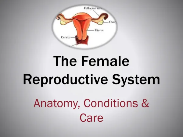
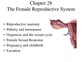
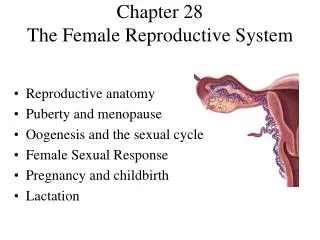
Chapter 28 The Female Reproductive System. Reproductive anatomy Puberty and menopause Oogenesis and the sexual cycle Female Sexual Response Pregnancy and childbirth Lactation. Female Reproductive System. Produce & deliver gametes Provide nutrition & room for fetal development Give birth

Chapter 28 The Female Reproductive System
E N D
Presentation Transcript
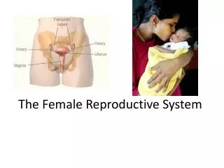
Chapter 28The Female Reproductive System • Reproductive anatomy • Puberty and menopause • Oogenesis and the sexual cycle • Female Sexual Response • Pregnancy and childbirth • Lactation
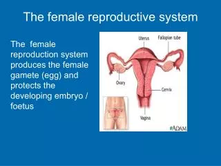
Female Reproductive System • Produce & deliver gametes • Provide nutrition & room for fetal development • Give birth • Nourish the infant
Sex Differentiation • Male & female are indistinguishable for the first 8 to 10 weeks of development • Female develops due to absence of hormones • absence of testosterone & müllerian-inhibiting factor causes degeneration of (male) mesonephric duct • phallus becomes clitoris, urogenital folds develop into labia minora & labioscrotal folds into labia majora • paramesonephric duct develops into uterine tubes, uterus and vagina
Ovary • Ovaries produce eggs & female hormones • almond-shaped organ, 3 cm x 1.5 cm x 1 cm • tunica albuginea capsule like the testes • cortex producing gametes & medulla holding vessels • Each egg develops in its own fluid-filled follicle & is released by ovulation, bursting of the follicle • Ligaments • attached to uterus by ovarian ligament • attached to pelvic wall by suspensory ligament • contains ovarian artery, vein & nerves • anchored to broad ligament by mesovarium
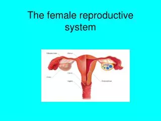
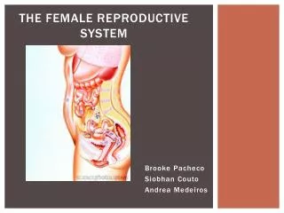
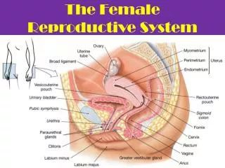
Secondary Sex Organs (Genitalia) • Internal genitalia • duct system consisting of uterine tubes, uterus & vagina • External genitalia • clitoris, labia minora, and labia majora • occupy the perineum • accessory glands beneath the skin provide lubrication
Uterine or FallopianTubes (Oviducts) • 10 cm long, muscular tube lined with ciliated cells • Major portions of tube • near uterus forms a narrow isthmus • middle portion is body (ampulla) • flared distally into infundibulumwith fimbriae • Enclosed in superior margin of broad ligament (mesosalpinx)
The Uterus • Thick-walled, pear-shaped muscular chamber opening into vagina and tilted forward over the urinary bladder • internal & external os of cervical canal • openings into uterine tubes in its two upper corners • Domed fundus above body of organ
Histology of the Uterine Wall • Perimetrium is external serosa layer • Myometrium is middle muscular layer • 1 cm thick in nonpregnant uterus • smooth muscle running in all directions • produces labor contractions to expel fetus during delivery • Endometrium • simple columnar epithelium with thick layer compound tubular glands • stratum functionalis is superficial 1/2 shed with each period • stratum basalis is deeper layer that regenerates a new stratum functionalis with each menstrual cycle
Vessels of Female Reproductive Tract • Hormonal changes cause spiral artery vasoconstriction, necrosis of the stratum functionalis & menstrual flow
Vagina or Birth Canal • 8-10 cm long, distensible muscular tube • allows for discharge of menstrual fluid, receipt of semen and birth of baby • Outer adventitia, middle muscularis & inner mucosa • in child, epithelium is simple cuboidal • estrogens of puberty transform into stratified squamous • bacteria ferment glycogen rich cells producing acidic pH • Tilted posteriorly between rectum & urethra • urethra embedded in its anterior wall
The Vulva (Pudendum) • Mons pubis = mound of fat over pubic symphysis • Labia majora = thick folds of skin (pubic hair) • Labia minora = more medial, thin hairless folds • form vestibule containing urethral & vaginal openings • form hoodlike prepuce over clitoris • Clitoris = erectile, sensory organ • homologous to glans penis of male • Vestibular bulbs = erectile tissue around vagina • Paraurethral and greater & lesser vestibular glands open into vestibule for lubrication
The Breasts • Mound of tissue overlying the pectoralis major • conical body of breast has nipple at its apex • axillary tail in armpit contains many lymphatic vessels • Nipple is surrounded by areola (colored zone) • dermal blood vessels are closer to surface • melanocytes darken during pregnancy • smooth muscle contracts wrinkling the skin & erecting the nipple in response to cold, touch & arousal • Suspensory ligaments attach it to skin & muscle • If nonlactating, contains little glandular tissue just a system of branching ducts and fat tissue
Breast Cancer • 1 out of every 8 American women • Tumors begin with cells from mammary ducts • may metastasize by way of lymphatics • Symptoms may include palpable lump, skin puckering, skin texture & drainage from the nipple • Most breast cancer is nonhereditary • some stimulated by estrogen • Risk factors include aging, ionizing radiation, carcinogenic chemicals, alcohol, smoking & fat intake (70% lack risk factors)
Puberty • Begins at age 9 or 10 for most girls in the U.S. • Triggered by rising levels of GnRH which stimulate anterior lobe of pituitary to produce FSH & LH (follicle-stimulating & luteinizing hormone) • FSH stimulates follicles to secrete estrogen & progesterone • 2nd sex organs maturation, in height & width of pelvis • prepares uterus for pregnancy • Thelarche = development of breasts • Pubarche = growth of pubic & axillary hair, apocrine & sebaceous glands • Menarche = first menstrual period (age 12) • requires at least 17% body fat in teenager, 22% in adult • Female hormones secreted cyclically & in sequence
Climacteric and Menopause • Midlife change in hormone secretion accompanied by menopause (cessation of menstruation) • average age of 52 • Age related depletion of follicles means less secretion of estrogen & progesterone • atrophy of uterus, vagina & breasts • skin becomes thinner, bone mass declines, and risks of cardiovascular disease increase • hot flashes (sudden dilation of cutaneous arteries) occur several times a day • HRT = low dose estrogen & progesterone therapy
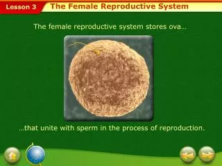
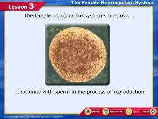
Oogensis and the Sexual Cycle • Reproductive cycle - events occurring between fertilization and birth • Sexual cycle - events recurring every month when pregnancy does not occur • ovarian cycle = events in the ovaries • menstrual cycle = parallel changes in the uterus
Oogenesis • Monthly event producing 1 haploid egg by meiosis • Embryonic development of ovary • female germ cells arise from yolk sac of embryo • differentiate into oogonia & multiply in number • transform into primary oocytes(eggs) -- early meiosis I • most degenerate (atresia) by time reach childhood • by puberty 400,000 oocytes remain • FSH stimulates completion of meiosis I, produces secondary oocyte & 1st polar body • proceeds to meiosis II & ceases until fertilization • after fertilization , releases 2nd polar body
Sexual Cycle • Averages 28 days but ranges from 20 to 45 • Hormone cycle produces hierarchy of control • hypothalamus pituitary ovaries uterus • Follicular phase (2 weeks) • menstruation occurs during first 3 to 5 days of cycle • uterus replaces lost endometrium & follicles grow • Postovulatory phase (2 weeks) • corpus luteum stimulates endometrial thickening • endometrium lost again if pregnancy does not occur
Ovarian Cycle -- Follicular Phase • From beginning of menstruation (day 1) to ovulation(14) • most variable part of cycle • Seldom possible to predict date of ovulation • Contains menstrual and preovulatory phases
Ovarian Cycle -- Menstrual Phase • During discharge of menstrual fluid (days 1-5) • The 25 primary oocytes that began developing on day 25 of previous cycle have been transformed into 2nd follicles by day 5 -- follicular fluid & corona radiata have formed
Ovarian Cycle -- Preovulatory Phase • From days 6 to 14, one follicle advances to graafian stage & protrudes from surface of ovary • atresia of other follicles occurs with FSH • Egg stopped at metaphase II stage of meiosis
Ovarian Cycle -- Ovulation • Results from a spike of LH(caused by estrogen from follicle) • blood flow causes follicle to swell rapidly; collagenase weakens ovarian wall; fluid oozes out with oocyte and is swept up into uterine tube by fimbriae
Ovarian Cycle -- Postovulatory Phase • Luteal phase - corpus luteum forms from ruptured follicle under direction of Luteinizing Hormone progesterone stimulates secretory phase of menstrual cycle (in uterus) • Premenstrual phase – if no pregnancy, corpus luteum corpus albicans progesterone menstruation
Menstrual Cycle -- Proliferative Phase • Time of rebuilding of endometrial tissue lost at last menstruation -- mitosis occurs in stratum basalis • Result of estrogen from developing follicles • Reaches 2-3 mm in thickness Proliferative phase
Menstrual Cycle -- Secretory Phase • Further thickening of endometrium due to secretion & fluid accumulation -- not mitosis • Due to progesterone stimulation of glands • Reaches 5-6 mm in thickness
Menstrual Cycle -- Premenstrual Phase • Progesterone level falls due to atrophy of corpus luteum • Spiral arteries constrict causing endometrial ischemia • Pools of blood accumulate in stratum functionalis
Menstrual Cycle -- Menstrual Phase • Blood, serous fluid and endometrial tissue are discharged • Average woman loses 40 mL of blood & 35 mL of serous fluid --- contains fibrinolysin so it does not clot
Pregnancy and Childbirth • Gestation (pregnancy) lasts an average of 266 days from conception to childbirth • Gestational calendar is measured from first day of the woman’s last menstrual period (LMP) • Birth is predicted to occur 280 days from LMP • 3 three month intervals called trimesters
Prenatal Development • Age based terminology • blastocyst is less than 2 weeks old • embryo is from 3 to 8 weeks old • fetus is 9 weeks to birth • newborn up to 6 weeks old is called a neonate • Blastocyst consists of inner cell mass (developing embryo) and outer cell mass (trophoblast) • implantation= attachment of conceptus to endometrium • placenta is both maternal & trophoblastic tissue • embryo attached to placenta by umbilical & floats in amniotic fluid
Hormones of Pregnancy (1) • HCG (human chorionic gonadotropin) • secreted by trophoblast within 9 days of conception • prevents involution of corpus luteum • Estrogens • increases to 30 times normal before birth • corpus luteum is source for first 12 weeks until placenta takes over • causes uterine, mammary duct & breast enlargement
Hormones of Pregnancy (2) • Progesterone secreted by placenta & corpus luteum • suppresses secretion of FSH & LH preventing follicular development • prevents menstruation & thickens endometrium • stimulates development of acini in breast tissue • HCS (human chorionic somatomammotropin) • called human placental lactogen • secreted from placenta in direct proportion to its size • mother’s glucose usage and release of fatty acids • Other endocrine organs • thyroid gland increases 50% in size BMR of mother • parathyroid glands enlarge & stimulate osteoclasts to release additional calcium from mother’s bones • Aldosterone secretion fluid retention & leads to in mother’s blood volume